Tổng số lượt xem trang

Thứ Sáu, 27 tháng 1, 2017

Shoulder Ultrasound often Changes Patient Management

January 16, 2017 -- Shoulder ultrasound findings frequently have a significant effect on management and clinician decision-making in patients being treated for shoulder pain, according to research from the Mallinckrodt Institute of Radiology (MIR) in St. Louis.
After retrospectively evaluating the impact of more than 900 shoulder ultrasound exams on patient management, the Mallinckrodt researchers found that nearly two-thirds of patients received a change in their treatment plan based on the ultrasound findings. What's more, shoulder ultrasound had a statistically significant effect on the invasiveness of the patient treatment.
Dr. Michael Friedman shared the MIR research during a session at RSNA 2016 in Chicago.
A common complaint
Shoulder pain is the third most common presenting musculoskeletal complaint after knee and low-back pain, affecting 16% to 26% of the population. It's a difficult clinical assessment, however, due to the overlap of many common shoulder pathologies, Friedman said.
Rotator cuff pathology is one of the most common causes of shoulder pain, for which MRI has been the gold standard. There's been a tremendous increase in the use of musculoskeletal ultrasound over the past five to 10 years, however. This growth is supported by research, such as a study that found 84% sensitivity and 89% specificity for partial-thickness tears and 96% sensitivity and 93% specificity for full-thickness tears, he said. MSK ultrasound also has been shown to have good correlation with MRI for rotator cuff muscular atrophy.
The Society of Radiologists in Ultrasound has published a consensus statement of recommended imaging algorithms for referring physicians on how to assess shoulder pain. Questions remain, however, about the actual diagnostic impact of musculoskeletal ultrasound on current orthopedic clinical evaluation of these patients.
As a result, the Mallinckrodt researchers set out to explore the effect of musculoskeletal ultrasound of the shoulder on clinical decision-making at their institution. A search of their RIS database found 1,037 consecutive shoulder ultrasound studies over a 12-month period, ordered by 15 orthopedic faculty members and interpreted by 10 radiologists. After 102 exams were excluded due to missing pre- and postultrasound clinical notes or because they were ordered for contralateral comparisons, the researchers were left with 935 patient exams for their study.
Three musculoskeletal radiologists then analyzed the electronic medical record and clinic notes and recorded the pre- and postultrasound treatment plans. Patients were classified into treatment plan categories, including conservative management/physical therapy (PT), therapeutic injection, or surgical intervention. Many patients were frequently prescribed conservative management/physical therapy and therapeutic injection together, which was also analyzed as a distinct management plan.
Conservative management/PT was considered to be noninvasive management, while therapeutic injection and surgical intervention were deemed to be invasive management.
Treatment changes
After receiving an ultrasound scan, 63.2% of patients had their treatment plans changed.
Patient management plans before and after MSK ultrasound
Preultrasound treatment plan
No. of patients
Overall change in management plan following MSK ultrasound
Conservative management/PT
744/935 (79.6%)
490/744 (65.9%)
Invasive management (injection or scheduled for surgery)
191/935 (20.4%)
101/191 (52.9%)
Of the 744 patients with an initial conservative management/PT treatment plan, 423 (56.9%) were shifted to a more invasive treatment plan based on the shoulder ultrasound exam. In patients set to receive an invasive management plan, 46 (24.1%) were changed to a noninvasive treatment plan.
The impact of shoulder ultrasound on the invasiveness of patient management was statistically significant (p < 0.001).

The researchers acknowledged the limitations of their study, including its reliance on the quality of physician documentation. The team did not make any assumptions about treatment intent and placed patients in treatment categories solely based on the impression in the clinical notes.

Thứ Ba, 24 tháng 1, 2017

TẠO HÌNH SWE CÁC HẠT GIÁP

TẠO HÌNH SWE CÁC HẠT GIÁP
V. CANTISANI et al

Hầu hết các máy siêu âm đàn hồi đo tốc độ sóng biến dạng bằng ROI nhỏ có kích thước chọn trước cố định đặt trong vùng muốn khảo sát. Kỹ thuật này được gọi là  điểm siêu âm đàn hồi p-SWE [SW elastography point] và cung cấp kết  quả số của SWS của mô trong ROI box cả bằng đơn vị  m/s hoặc kPa. Giá trị độ cứng cao tương ứng với tốc độ cao. Kỹ thuật ARFI là nhãn hiệu phần mềm đầu tiên dùng p-SWE.

Mặt khác, vài máy có khả năng hiển thị số đo ARFI mã hóa màu trong môt hộp ROI lớn, là bản đồ tốc độ, còn gọi là hình đàn hồi hay elastogram. Kích thước của elastogram do người đo kiểm soát bằng tay và hiển thị chồng lên hình B-mode, gọi là 2D-SWE.
Trên hình siêu âm đàn hồi có những vùng không màu có thể do chứa dịch, vôi hóa, hoặc  không thể đo do vấn đề kỹ thuật. Trên vài máy có hiển thị số đo là “X.XX” hoặc “0.00” vì hạt giáp chứa dịch hay vôi hóa.

Với p-SWE, đôi khi máy không đo chính xác do một số yếu tố vì chuyển động hoặc nhiễu, hoặc biên độ quá thấp để tạo sóng biến dạng, hay hạt giáp quá cứng nên tốc độ biến dạng quá cao không thể lấy mẩu. Chẳng hạn như ARFI khi không thể đo sẽ hiển thị “X.XXm/s”, khi đó chỉ cần đổi vị trí đầu dò và đo lại.

GIÁ TRỊ CHẨN ĐOÁN SWE CÁC HẠT GIÁP:


Theo y văn , đối với p-SWE tốc độ sóng biến dạng của tuyến giáp bình thường là 1, 60 ± 0,18 m/s. Với 2D-SWE dùng SSI là 2,6 ± 1,8 m/s và 20,8 ± 10,4 kPa . Phân biệt hạt giáp lành với ác tính, SWE có giá trị ngưỡng từ 3,65 m/s to 4,70 m/s (34, 5–66 kPa).

 Tốc độ sóng biến dạng của tổn thương ác tính cao hơn lành tính có ý nghĩa, với giá trị cao hơn 2,87m/s gợi ý ác tính. Tất cả nghiên cứu và phân tích hồi cứu cho rằng SWE (p-SWE và 2D-SWE) có thể là phương tiện bổ sung có ích giúp phân biệt hạt giáp lành tính và ác tính.

Dịch từ Atlas of Elastosonography, D-A CLEVER et al, © Springer Intern Publ. Switzerland 2017.
----


 How to Improve the Examination

1. In all SWE techniques, for accurate measurements, a slight contact of the probe to the skin is essential, as pressure applied by the probe alters the measurements and causes artifacts of increased stiffness. For this reason experience is required to perform reliable examinations and SWE quantification. Liberal use of ultrasound coupling gel can help maintain only slight contact with the skin. The patient should perform adequate breath hold.
2. If you encounter a pattern of red in the near field, also called a vertical artifact, you may reduce it by minimizing probe compression.

3. The isthmic nodules, compressed against the trachea, are the most at risk to effect of probe pressure. To avoid the artifactually increased measured stiffness, some authors advise the use of paracoronal scanning in order to avoid compressing the nodules against the trachea
.
4. The ROI should include as much as possible of the area of the nodule. Normal thyroid parenchyma should not be included in the ROI.
5. ROI must be placed in the nodule avoiding cystic or calcified areas as these alter the measurements. However, the use of the standard size ROI in p-SWE sometimes makes it impossible to exclude fluid and calcified areas of a nodule.
6. Choosing a correct preset is important to acquire accurate measurements, and the preset should be 0–180 kPa for the thyroid application
 Increase the elasticity color gain to its maximum till the noise starts to appear in the elasticity image, because this will aid in acquiring good color sensitivity elastograms.
7. With 2D-SWE in order to obtain the most significant quantitative readouts, ROIs should be placed on the stiffest part of a lesion as shown in the velocity map (2D-SWE elastogram). Malignancy is often very heterogeneous in stiffness; it is useful to choose the stiffest site for the measurement.
8. In the elasticity image, there can be areas with no color that may be due to fluid content, calcification, or impossible velocities’measurements due to technical problems
. In some systems the displayed measurements show “X.XX” or “0.00” because these nodules contain cysts and calcifications. Cysts, when containing nonviscous fluids, do not support shear waves, and so they appear as color voids, usually seen as black regions where the anechoic B-mode layer shows through. On the other hand, when the cyst fluid is viscous, shear wave signals may be seen and would display a soft region.
9. With p-SWE, sometimes the system cannot calculate an accurate measurement due to a variety of factors such as motion or noise or probably a too low amplitude of the generated shear wave or even too high stiffness in the nodule with a too high shear wave speed that cannot be sampled
. ARFI for example, when it cannot  yield a measurement, displays “X.XX m/s” instead of the expected numeric value. In such an occurrence,slightly change the position of the probe and repeat the evaluation.
10. With ARFI, in the experience of some researchers, ten measurements were needed to obtain an average which yielded a reliable accuracy. Another research group had good results with five measurements.
11. Always bear in mind that, as with strain elastography, malignancy may appear genuinely soft, albeit rarely. Follicular carcinomas, in particular, can be soft and difficult to distinguish from benign nodules.
12. For good documentation, some authors recommend, for every nodule, the acquisition of three cine loops, each of at least 10 s of duration
.

SIÊU ÂM ĐÀN HỒI TINH HOÀN

SIÊU ÂM ĐÀN HỒI TINH HOÀN
Dirk-Andre – Clevert

Siêu âm là kỹ thuật nhạy và chính xác trong khảo sát các bất thường của tinh hoàn, được chấp nhận là tiêu chuẩn vàng dù không cung cấp chẩn đoán mô học.
Do phát triển với những đầu dò HF, siêu âm màu Doppler và real-time elastography, siêu âm được xem như là kỹ thuật CĐHA được chọn cho khám phần mềm và bệnh lý bìu.
Real–time elastography hay đàn hồi căng đã được triển khai hơn 20 năm nay khảo sát không xâm lấn đặc điểm cơ học của mô và tạo hình đàn hồi mô sinh học, trong đó có đánh giá các khối u ở bìu. Tuy nhiên, RTE chỉ định tính hay bán định lượng, lại tùy thuộc người khám nên phải đối mặt với những mức độ biến thiên.

VTIQ Siemens là kỹ thuật mới đo độ cứng mô real-time, sử dụng ARFI tự động tạo nên sóng biến dạng trong mô đích. VTIQ không dựa vào áp lực cơ học trên mô của người khám, và vượt qua các trở ngại truyền thống của đàn hồi căng bằng cách cung cấp các thông tin định lượng như tốc độ truyền sóng biến dạng trong mô đo bằng m/s và hiển thị bằng bản đồ mã màu 2 chiều. Tốc độ sóng biến dạng càng cao, độ cứng mô càng được khảo sát. Tuy nhiên vì shear waves không thể hiện các  biến dạng dịch, các nang tinh hoàn không đo được như khảo sát của Barr và Trottmann cùng cs.

Dịch từ Atlas of Elastosonography, D-A CLEVER et al, © Springer Intern Publ. Switzerland 2017.

Thứ Hai, 23 tháng 1, 2017

SIÊU ÂM ĐÀN HỒI ĐỘNG MẠCH CẢNH

SIÊU ÂM ĐÀN HỒI ĐỘNG MẠCH CẢNH
DIRK-ANDRE CLEVERT

Stroke là nguyên nhân gây chết thứ ba sau nhồi máu cơ tim và ung thư.
Xơ vữa động mạch có thể tác động chính mạch máu nội sọ; 88% bệnh nhân mù đột ngột do lấp mạch [amaurosis fugax] hoặc tai biến thiếu máu bán cầu não thoáng qua đều có bệnh vữa xơ động mạch ở chỗ chia đôi động mạch cảnh.
Vào khoảng 25-50% các loại tai biến là do mảng xơ vữa động mạch cảnh mất ổn định và khoảng 80% các tai biến là thiếu máu não.
Điều trị hẹp động mạch cảnh trong cần phân biệt chẩn đoán trước can thiệp là hẹp mức độ cao, giả tắc và tắc.
Những kết quả của  NASCET và ECST cho thấy bênh nhân hẹp mức độ cao 70-90% động mạch cảnh trong  có triệu chứng được lợi khi làm carotid endarterectomy [CEA], theo guidelines của AHA và EC của ACAS, CEA cũng là phương thức được chọn để điều trị bệnh nhân hẹp  ICA mức cao không triệu chứng.
Tuy nhiên khi làm surgical endarterectomy hoặc đặt stent động mạch cảnh để làm giảm nguy cơ stroke, có 3-9% bệnh nhân qua can thiệp này phải bị stroke hoặc chết do biến chứng điều trị. Bệnh nhân đặt stent có  biến chứng nguy cơ cao hơn làm endarterectomy.
Phương pháp đánh giá bệnh lý động mạch cảnh không xâm lấn là Doppler màu và phổ cho đoạn ngoài sọ. Nhưng Doppler có các hạn chế và không thể đánh giá đầy đủ khi phân biệt hẹp nặng với tắc hoàn toàn khi Duplex Doppler không tối hão.
Hiện nay chỉ có vài bài báo được in về phân tích đo đạc  tissue strain trong bệnh lý mạch máu. 


Dịch từ Atlas of Elastosonography, D-A CLEVER et al, © Springer Intern Publ. Switzerland 2017.

ĐỌC THÊM:

Shear Wave Elastography May Be Superior to Grayscale Median for the Identification of Carotid Plaque Vulnerability: A Comparison with Histology, F. Ozkan, D. Ozel, B. D. Ozel


... The authors report that shear wave elastography (SWE) findings regarding plaque stiffness may be used to help identify vulnerable plaque [1]. However, they found that there is a large overlap in Young’s Modulus values between stable and unstable plaques and this indistinctness requires attention to the interpretation of the ultrasound measurements for evaluating patients in routine clinical practice.
Because of the prominent role of carotid plaque in stroke incidence, their biomechanics have attracted significant scientific interest. There are several reports in the literature regarding the use of different methods to determine properties of plaque components [2] [3] [4]. In these methods, viscoelasticity is one of main parameters for clearly characterizing atherosclerotic plaques [2] [3] [4]. Elastography imaging is a novel ultrasound technique for quantifying tissue elasticity. However, studies have generally focused on the elastic properties of tissue, not on the viscosity [5]. Moreover, it has been claimed that if viscosity is not regarded, measurements of stiffness can be biased, and the results may be greater than the real values [6] [7]. Therefore, it is essential that viscosity be measured in order to fully characterize atherosclerotic plaques.
Garrard et al. could easily have analyzed the viscosity parameters of the atherosclerotic plaques because in a recent study, the viscosity in the liver was measured using post-processing techniques and the same ultrasound imaging system as Garrard et al. (Aixplorer, Supersonic Imagine, Aix-en-Provence, France) [8]. We believe that the large overlap in Young’s Modulus might be decreased by adding viscosity measurements.

Shear wave elastography imaging of carotid plaques: feasible, reproducible and of clinical potential

Kumar V Ramnarine, James W Garrard , Baris Kanber , Sarah Nduwayo , Timothy C Hartshorne and Thompson G Robinson

Abstract

Background: Shear Wave Elastography (SWE) imaging is a novel ultrasound technique for quantifying tissue elasticity. Studies have demonstrated that SWE is able to differentiate between diseased and normal tissue in a wide range clinical applications. However its applicability to atherosclerotic carotid disease has not been established.
The aim of this study was to assess the feasibility and potential clinical benefit of using SWE imaging for the assessment of carotid plaques.

Methods: Eighty-one patients (mean age 76 years, 51 male) underwent greyscale and SWE imaging. Elasticity was quantified by measuring mean Young’s Modulus (YM) within the plaque and within the vessel wall. Echogenicity was assessed using the Gray-Weale classification scale and the greyscale median (GSM).

Results: Fifty four plaques with stenosis greater than 30% were assessed. Reproducibility of YM measurements, quantified by the inter-frame coefficient of variation, was 22% within the vessel wall and 19% within the carotid plaque. Correlation with percentage stenosis was significant for plaque YM (p = 0.003), but insignificant for plaque GSM (p = 0.46). Plaques associated with focal neurological symptoms had significantly lower mean YM than plaques in asymptomatic patients (62 kPa vs 88 kPa; p = 0.01). Logistic regression and Receiver Operating Characteristic (ROC) analysis showed improvements in sensitivity and specificity when percentage stenosis was combined with the YM (area under ROC = 0.78).

Conclusions: Our study showed SWE is able to quantify carotid plaque elasticity and provide additional information that may be of clinical benefit to help identify the unstable carotid plaque.

SIÊU ÂM ĐÀN HỒI THẬN

SIÊU ÂM ĐÀN HỒI THẬN
Costanza Bruno et al

Ngay từ đầu, siêu âm đàn hồi thận là thách thức vì cho nhiều kết quả khác nhau. Đầu mối tìm hiểu các xung đột kết quả trên có thể từ phân tích nhiều yếu tố ảnh hưởng đến việc đo đàn hồi ARFI. Nhiều kết quả thì do mô đích khác nhau trong khi, quan trọng nhất là liên quan với cấu tạo giải phẫu thận.

Thận là cơ quan không đẳng hướng cao, gây ra những hồi âm theo các hướng tùy thuộc vào vài đặc tính của các thành phần cấu tạo. Đặc biệt, tủy thận [nơi các quai Henle và các ống thẳng song song nhau được hướng từ bao đến nhú thận] rất khác biệt với vỏ, hầu như chiếm bởi các cầu thận và các ống lượn gần và xa nên có dạng hình cầu. Điều này có nghĩa là khi chùm siêu âm song song đến với một phần thận, SW tạo ra sẽ đi vuông góc với các cấu trúc tủy thận, do vậy tạo ra vô số các mặt phẳng phân cách, và do vậy tốc độ lan truyền giảm xuống. Ngược lại, khi chùm siêu âm vuông góc với các quai Henle và các ống thẳng, SW tạo ra sẽ truyền đi song song với các cấu trúc này và ít gặp các mặt phẳng phân cách hơn; điều này sẽ đo SW cao hơn phản hồi, như trong một mô hình thực nghiệm của Supersonic Shear Wave.

Đàn hồi, biến thiên bình quân của shear modulus biểu kiến do không đẳng hướng tủy thận là 31,8% tương đồng với 40% của phân đoạn không đẳng hướng khi dùng diffusion MRI. Ngoài ra, không đẳng hướng của các yếu tố khác có liên quan chặt với chức năng của chúng có thể gây ảnh hưởng đánh giá thận bằng ARFI.
Trong cùng một mô hình nghiên cứu, đã chứng minh có tương quan tuyến tính giữa tăng áp lực niệu do thắt niệu quản. Thay đổi dòng chảy động mạch và tĩnh mạch cũng được tìm thấy gây ảnh hưởng điều hòa SWV; đặc biệt tắc động mạch thận gây giảm và thắt tĩnh mạch thận gây tăng.
 Các kết quả tương tự cũng được tìm thấy trong mô hình thực nghiệm khác, với 2 điều kiện khác nhau: tắc niệu quản và tắc tĩnh mạch thận. Mức độ hóa xơ khác biệt có ý nghĩa khi so sánh với thận bình thường; hóa xơ do gây tắc niệu quản thấp hơn hóa xơ do gây tắc tĩnh mạch thận. SWV có tương quan dương tính với biểu hiện mô học.
Bên cạnh những yếu tố này  thuộc về cơ quan đích, còn có những yếu tố khác ảnh hưởng đến truyền SW, đặc biệt là đè ép thận, khi đánh giá các thận ghép nông. Do đó phải sử dụng kỹ thuật chuẩn và chính xác nhằm làm giảm biến thiên khi đo.
Vì ứ đọng nước tiểu bàng quang có ảnh hưởng đến dữ liệu nghiên cứu, nên khảo sát sau khi đi tiểu, và vùng ROI song song với chùm siêu âm chính và hoàn toàn trong vùng vỏ thận (khó khi bề dày vỏ thận giảm đi và ROI cố định); và để làm giảm tình trạng không đẳng hướng,  và  với để cùng trục với tháp thận, nên chọn làm ở 1/3 giữa thận là dễ dàng nhất.


Về tương quan giữa tần số đầu dò và SWV, nên dùng HF probe 6 MHz cho trẻ em và cho người lớn 4 MHz, để có những kết quả có thể đối chiếu.
Không có đồng thuận nào về tư thế, có tư thế nằm sấp, nghiêng bên và nằm ngữa.
ARFI ở trẻ em thường thấp và tăng dần theo tuổi, chú ý trẻ dưới 5 tuổi.

BỆNH LÝ THẬN LAN TỎA
Sau những kêt quả ARFI đạt được trong đánh giá gan, xu hướng đánh giá thận được phát triển giả định dựa vào tế bào to, xơ hóa kẽ mức độ cao dẫn đến mất chức năng nephron, là nguyên nhân bệnh lý thận mạn tính.
Tiếp cận không xâm lấn bệnh lý thận mạn tính cần cả các thông số hình thái [kích thước, bề dày vỏ thận, đường kính bể thận] và chỉ số cản RI giúp định lượng và xác định mức độ tổn thương. Điều này giúp sinh thiết thận nếu cần và chụp scintigraphy với 99mTc-DMSA theo guidelines trong đánh giá vài dạng của trẻ em.

Thận ghép:
Kết quả rất khác nhau
Trong nghiên cứu thăm dò, Amdt cho thấy độ cứng chủ mô đánh giá bằng TE có liên quan với xơ hóa mô kẽ, có tương quan tốt giữa độ cứng vỏ với thoái hóa mô toàn bộ ở Supersonic SWE.
He và cs thấy có tương quan giữa SWV và eGFR với độ nhạy 72% và độ chuyên biệt 86,5% trong đánh giá mất chức năng thận ghép. Thận ghép bị thải ghép cấp có SWV thấp hơn thận ghép có bệnh lý thận, giá trị SWE trung bình cao hơn  ở bệnh nhân có eGFR thấp hơn [<50 vs=""> - 50mL/min].
Mặt khác Syversveen và cs nhận thấy ARFI không thể phát hiện xơ hóa thận ghép với xu hướng giảm SWV trung bình từ xơ hóa độ thấp đến cao và tương tự như  Gao và cs báo cáo, giá trị độ cứng cao hơn ở bn xơ hóa nhỏ hơn 25% hơn ở nhóm xơ hóa lớn hơn 26%.


Thận mạn tính:
Vì cho đánh giá thận ghép mạn tính cũng như xem xét bệnh thận mạn, kết quả rất khác nhau do vấn đề chuẩn hóa nghiên cứu.
Wang báo cáo không tương quan giữa mức độ xơ hóa với SWV ở 45 bn, phần đông bị bệnh lý thận IgA. Các tác giả khác nhận thấy SWV giảm dần với sự tăng CKD và với giảm GFR. Điều này có thể giải thích do giảm dần lưu lượng máu thận do xơ hóa mạch máu thận, làm ảnh hưởng đến SWV hơn là mô thận xơ hóa. Kết quả tương tự xảy ra ở bệnh nhân thận mạn tính do tiểu đường. ARFI có khả năng phân biệt các giai đoạn bệnh thận tiểu đường khác nhau; có tương quan đảo ngược giữa giá trị ARFI và GFR.
Phát hiện này bao gồm phát hiện của thực nghiệm của Gennisson cho thấy SWV giảm sau thắt động mạch thận.

Thận trẻ em:
Đánh giá thận trẻ em là thách thức đặc biệt do trẻ em khó nín thở khi khám. Ứng dụng chính là khảo sát bệnh thận mạn và xử trí tổn thương thận khi thận ứ nước.
Trẻ dưới 2 tuổi với bể thận giãn, SWV cao hơn với ứ nước thận mức độ cao khi so với thận bình thường, mặc dù ARFI không phân biệt được ứ nước thận bế tắc với ứ nước thận không do bế tắc.
Dù có liên quan giữa loại bế tắc và SWV, trẻ em với bệnh lý thận mạn tính do bế tắc có ARFI cao hơn trẻ bị thận mạn tính không do bế tắc. Ở trẻ bị bệnh một bên, thận bên lành lại có SWV cao hơn nhóm chứng.


Bệnh lý khu trú:

Các báo cáo đầu tiên của  ARFI đánh giá khối u thận đã công bố và đặc biệt phân biệt lành và ác tính. Sau những cố gắng ban đầu đánh giá các u thận để thu thập các thông tin về bản chất tổn thương, nhiều nghiên cứu chuẩn được thực hiện.
Nang thận có SWV thấp.
Khác biệt giữa RCC và AML có ý nghĩa thống kê trong một cohort study số liệu lớn với điểm cut off là 1,87m/s với độ nhạy 47,5% và độ đặc hiệu là 33,2%. Oncocytoma thường gây ra vấn đề khi phân biệt với RCC, thường cũng nhiều mạch máu, có SWV thấp hơn.
Khi khảo sát khác biệt của nhóm phụ của RCC, SWV của clear cell RCC cao hơn SWV của papillarry RCC, chromophobe RCC và angiomyolipoma AML.
Với kích thước ROI cố định 1x0,5 cm, đặc biệt khó đánh giá một khối không đồng nhất hoặc một tổn thương dạng nang, vì cần tránh các vùng hoại tử hay nang hóa để có kết quả có thể lập lại.

Trích dịch từ Atlas of Elastosonography, D-A CLEVER et al, © Springer Intern Publ. Switzerland 2017.

Chủ Nhật, 22 tháng 1, 2017

SIÊU ÂM ĐÀN HỒI LÁCH

SIÊU ÂM ĐÀN HỒI LÁCH

Trích dịch từ Atlas of Elastosonography, D-A CLEVER et al, © Springer Intern Publ. Switzerland 2017.

Ứng dụng chính của đàn hồi lách nhằm đánh giá bệnh nhân viêm gan mạn để xác định nguy cơ cao áp tĩnh mạch cửa. Các nghiên cứu gần đây đã chứng minh độ cứng lách liên quan với hóa xơ gan, cao áp tĩnh mạch cửa và nguy cơ giãn tĩnh mạch thực quản ở bệnh nhân viêm gan mạn.
Cao áp tĩnh mạch cửa là hậu quả chung của bệnh gan mạn tính, dẫn đến giãn tĩnh mạch thực quản và dạ dày và các biến chứng nặng khác như bệnh lý não do thông nối cửa và hệ thống, báng bụng, nhiễm trùng và suy gan.
Tiêu chuẩn vàng của đánh giá cao áp tĩnh mạch cửa là gradient áp lực tĩnh mạch gan (HVPG, hepatic venous pressure gradient) mà không may là thủ thuật có tính xâm lấn và tốn kém nên không thường được triển khai theo quy trình ứng dụng lâm sàng. Cho nên siêu âm đàn hồi lách là một trong những phương pháp không xâm lấn, khả dĩ xác định áp lực tĩnh mạch cửa. Cao áp tĩnh mạch cửa xác định những thay đổi mô học trong lách như tăng sinh mô bào lách, kéo dài ra các nhánh tận động mạch, tăng thể tích chồi trắng và ngay cả hóa xơ giữa các bè lách. Những thay đổi này gây tăng đàn hồi lách và có thể định lượng bằng transient elastography (TE) hay ARFI.

Một nghiên cứu mới đây chứng minh đo đàn hồi gan và lách bằng TE chính xác hơn các thông số khác, và gợi ý dùng TE như là phương tiện đánh giá không xâm lấn và theo dõi cao áp tĩnh mạch cửa và giãn tĩnh mạch thực quản. Mặc dù TE thường dùng để phân độ bệnh gan mạn tính, có bằng chứng cho thấy sự chính xác chẩn đoán chưa phải tối ưu, do nhiều yếu tố cộng tồn như viêm tế bào gan, ứ mật và nhiễm mỡ làm cho đánh giá hóa xơ gan chưa chính xác. Do vậy, việc đo độ cứng lách cho thấy là một thách thức hấp dẫn, có thể làm giảm các yếu tố cộng tồn TE gan đã nói và tăng cường đánh giá độ nặng của bệnh.
Độ đàn hồi lách có tương quan tuyến tính chặt với HVPG, và có thể giúp tiên đoán giãn tĩnh mạch thực quản ở bệnh nhân viêm gan mạn. Giãn tĩnh mạch thực quản hiện diện ở 40% ở bệnh nhân còn bù và 60% bệnh nhân xơ gan mất bù. Và nguy cơ chính là xuất huyết tiêu hóa nặng (tỉ lệ tử vong của cơn XHTH đầu của những bệnh nhân này là 40%). Esophagogastroduedenoscopy (EGD) là phương tiện chẩn đoán và đánh giá giãn tĩnh mạch thực quản và dạ dày tốt nhất; tuy nhiên rất tốn kém cho hệ thống y tế và bệnh nhân khó chịu. Nên hạn chế cho những ca thực sự cần thiết và điều này làm tăng tin cậy cho bệnh nhân có quy trình theo dõi và giảm nguy cơ do biến chứng can thiệp.

Những nghiên cứu gần đây chứng minh độ cứng lách cao hơn độ cứng gan, chiều dài lách và thang điểm Child-Pugh trong tiên đoán xuất huyết giãn tĩnh mạch, cho thấy ARFI có độ nhạy 96%, và độ đặc hiệu 88% và giá trị tiên đoán âm tính là 96%.
Tuy nhiên, những nghiên cứu này cho thấy sự lập lại kết quả đo độ cứng lách ở những dân số khác nhau tùy thuộc vào người khám, và để có đồng thuận cao, cần có giai đoạn huấn luyện. Bất lợi khác là tương quan của những đánh giá gián tiếp. Nên thực hiện những nghiên cứu tiếp sau với số lượng lớn hơn để so sánh giá trị đàn hồi bình thường và mô bệnh lý để xác dịnh vai trò chẩn đoán của những kỹ thuật này. Cuối cùng, đo độ cứng lách bằng siêu âm có được dùng trong chức năng TIPS [transjugular intrahepatic portosystemic shunt] ở bệnh nhân viêm gan mạn. Đo độ cứng lách có thể là phụ trợ sau khi đặt TIPS, và sự thay đổi này có thể giúp đánh giá thay đổi chức năng của TIPS.


Có ít thông tin đáng giá  về sử dụng siêu âm đàn hồi các khối lách khu trú.


Thứ Hai, 9 tháng 1, 2017

LIVER ELASTOGRAPHY= EFSUMB GUIDELINES


LINK    DOWNLOAD  Ultrasound Med Biol. 2015 May;41(5):1161-79. doi: 10.1016/j.ultrasmedbio.2015.03.007. Epub 2015 Mar 20


....

POINT SHEAR WAVE SPEED MEASUREMENT (PSWSM) AND SHEAR WAVE SPEED IMAGING (SWSI)

Procedure

All technologies are implemented in a conventional US system under direct visualization using a curved array broadband transducer. A sample box is positioned on B-mode image of the liver and elastography measurements are obtained by pressing a button. Optimal conditions include:
 -Fasting;
 -Dorsal decubitus position, with the right arm elevated above the head for optimal intercostal access;  
 -Resting respiratory position (breath-hold without deep inspiration);  
 -ROI placement beneath Glisson’s capsule by 1.5-2.0 cm to avoid reverberation artifacts and increased subcapsular stiffness;
 - ROI placement to avoid large liver vessels;  
 -The median value of 5-10 measurements is considered with PSWSM, and the mean value of 4 measurements with SWSI.

Specific recommendations include:  

+For SWSI, the sample box size should be large enough to reduce the variation between measurements. This provides a cumulative value that is the average of stiffness at several points, thus being more representative of the heterogeneous stiffness in abnormal and normal livers.  
+For PSWSM, the ROI should be placed perpendicular to the center of the transducer surface as the angle of insonation may have a slight but significant influence on the result.

Results
Point Shear Wave Speed Measurement.

As of today, there are two techniques: Virtual Touch Tissue Quantification (VTTQ) technique that expresses the results in m/sec (Figure 3) and ElastPQ that gives the results in m/sec or in kPa (Figure 4). There are numerous reports of studies performed using the VTTQ technique, which has been commercially available since 2009, but only a few using ElastPQ, which was introduced in 2012. The reproducibility of the VTTQ technique is excellent, with an intraclass correlation coefficient ranging from 0.84 to 0.87 (Bota et al. 2012; Boursier et al. 2010; D’Onofrio et al. 2010; Guzman-Aroca et al. 2011). Operator training does not appear to be necessary (Boursier et al. 2010). Similarly, the ElastPQ technique is highly reproducible, with an interobserver agreement ranging from 0.83 for comparison of single measurements to 0.93 for the median value of 10 measurements (Ferraioli et al. 2014). In healthy volunteers, the values of PSWSM performed with VTTQ are available in several publications (D’Onofrio et al. 2010; Friedrich-Rust et al. 2009a; Goertz et al. 2012; Grgurevic et al. 2011; Kaminuma 2011; Karlas 2011; Kim 2010; Kircheis 2012; Osaki 2010; Piscaglia 2011; Rifai 2011; Rizzo et al. 2011; Son et al. 2012; Sporea et al. 2011; Takahashi et al. 2010). In all studies, the values were lower (, 1.2 m/sec) than in patients with chronic hepatitis. Food intake significantly increases the liver stiffness values (Goertz et al. 2012; Popescu et al. 2013). The median value of PSWSM obtained with ElastPQ in healthy volunteers is 3.5 kPa (Ling et al. 2013; Ferraioli et al. 2014).

Chronic viral hepatitis.
 The range of cut-offs for each fibrosis stage is quite large with overlap between consecutive stages. The range of cut-offs for the fibrosis stage ranges from 1.13 to 1.55 m/sec for F.2; from 1.43 to 1.81 m/sec for F.3; and from 1.36 to 2.13 m/sec for F4. The largest series comprises more than 600 patients with mixed etiologies of chronic liver disease (Kircheis et al. 2012). Using TE as the reference method, the investigators obtained cut-off values of 1.32 m/sec for F2 and 1.62 m/sec for F4. Similar cut-offs were obtained in the meta-analysis of Friedrich-Rust et al. (2012a), in which nine studies were analyzed. Patients with chronic liver disease of several etiologies were included, and the cut-off values were 1.34, 1.55 and 1.80 m/sec, for significant fibrosis, severe fibrosis and cirrhosis, respectively. PSWSM showed accuracy similar to that of TE for the diagnosis of severe fibrosis, whereas a slightly but significantly higher diagnostic accuracy of TE with respect to PSWSM was found for the diagnosis of significant fibrosis and liver cirrhosis. In the more recent metaanalysis of Bota et al. (2013), in which thirteen studies were included, PSWSM showed a predictive value similar to TE for significant fibrosis and cirrhosis. In an international multicenter study comprising 1,095 patients (181 with chronic hepatitis B and 914 with chronic hepatitis C), the correlation of PSWSM with histological fibrosis was significantly higher in patients with chronic hepatitis C compared with those with chronic hepatitis B (r50.653 vs. r50.511, p50.007), whereas both groups showed similar PSWSM values for each fibrosis stage (Sporea et al. 2012). In the study of Rizzo et al. (2011), using the PSWSM cut-offs of 1.3 m/sec for the diagnosis of significant fibrosis (F $ 2), 1.7 m/s for severe fibrosis (F $ 3), and 2.0 m/sec for cirrhosis (F 5 4), the highest concordance was obtained for the diagnosis of mild fibrosis. TE may overestimate the fibrosis stage in cases with severe liver inflammation (Sagir et al. 2008; Arena et al. 2008). The same limitation has been observed in some studies with PSWSM (Takahashi et al. 2010; Yoon et al. 2012; Chen et al. 2012), but not in others (Friedrich-Rust et al. 2009a; Palmeri et al. 2011; Rizzo et al. 2011; Nishikawa et al. 2014).


 Figure 3. VTTQ technique in a healthy subject. Measurements of liver stiffness are given in m/sec; the sample box is shown.


Figure 4. ElastPQ in a patient with chronic hepatitis C of F4 Metavir stage on liver histology. The values of liver stiffness are expressed in kPa. Bottom left corner of the image: the stiffness is estimated and displayed by using a scale that ranges from soft to hard.

The grade of liver steatosis appears not to influence PSWSM (Friedrich-Rust et al. 2009a; Rizzo et al. 2011; Rifai et al. 2011). Measurement failure with PSWSM is reported in less than 3% of patients (Friedrich-Rust et al. 2012a). No invalid measurement occurred in the series of Rizzo et al. (2011) and in the study of Crespo et al. (2012). PSWSM provided valid results in all patients whereas TE failed in 11% of cases. In the series of Bota et al. (2014), reliable measurements were obtained in 93.3% of cases. Older age, higher BMI and male gender were associated with the risk of failed and unreliable measurements. A recent meta-analysis, which included either full papers or abstracts for a total of 36 studies, has shown that BMI has a significant influence for the diagnosis of significant fibrosis (F $ 2) (Nierhoff et al. 2013). In this meta-analysis, the diagnostic accuracy expressed as the area under the ROC curve was 0.84, 0.89 and 0.91 for the diagnosis of significant fibrosis, advanced fibrosis, and cirrhosis, respectively. Measurements are not limited by ascites because the US push beam, which generates the shear waves, propagates through fluids and appears not to be influenced by clinical and biochemical variables (Rizzo et al. 2011). The possibility to evaluate several areas of the liver parenchyma could be another advantage of PSWSM. Indeed, histological studies have shown that liver fibrosis is not homogeneously distributed within the liver, and can be missed when performing liver biopsy at one site only, thus leading to underestimation of liver fibrosis (Bedossa et al. 2003; Maharaj et al. 1986). It should be noted that D’Onofrio et al. (2010) reported significant differences between intercostal and subcostal scans, and Kaminuma et al. (2011) found that PSWSM were more reliable when performed in a deep portion of the right lobe. In their series of patients with chronic hepatitis, Toshima et al. (2011) obtained significantly higher values in the left lobe of the liver than the right lobe. It has been suggested that oscillation of the left liver by cardiac activity may interfere with stiffness measurements (Osaki et al. 2010). Karlas T et al. (2011) found that, in healthy individuals, the shear wave speed was higher in the left liver than in the right, but no difference in speed was observed in patients with advanced fibrosis and cirrhosis. The authors suggest that the absence of differences between the two sides could be a criterion for the diagnosis of advanced liver disease. Preliminary results of PSWSM using ElastPQ in 102 patients with chronic hepatitis C have shown that the accuracy of the method for staging liver fibrosis is similar to that of TE and the best cut-off value for significant fibrosis (F $ 2) is 5.7 kPa (Ferraioli et al. 2014). In a series of 291 patients with chronic hepatitis B, the AUROCs for significant fibrosis and cirrhosis were 0.94 and 0.89, respectively (Ma et al. 2014).

Monitoring disease progression and prognosis.
Very few studies regarding disease progression and prognosis have been published with conflicting results. In the cohort of Vermehren et al. (2012), the diagnostic accuracy of PSWSM of the liver and the spleen for the prediction of esophageal varices was not signifi- cantly different from that of TE and the Fibrotest; however, the AUROCs of all methods were fairly low, ranging from 0.50 to 0.58. In the series of Morishita et al. (2013), a cutoff value of 2.39 m/s had a sensitivity of 81% and a specificity of 82% for detecting high-risk esophageal varices. In a recent study, a spleen stiffness value ,3.3 m/s ruled out the presence of high-risk varices in patients with compensated or decompensated liver cirrhosis (negative predictive value, 99.4%). Regardless of the etiologies of liver disease, spleen stiffness was highly accurate for the detection of esophageal varices (Takuma et al. 2013).

 Nonalcoholic fatty liver disease (NAFLD).
Only few studies in small series of patients are available (Yoneda et al. 2010; Osaki et al. 2010; Palmeri et al. 2011; Friedrich-Rust et al. 2012b; Fierbinteanu Braticevici et al. 2013). In 172 patients diagnosed with NAFLD, a cutoff of 4.24 kPa distinguished low (fibrosis stage 0–2) from high (fibrosis stage 3–4) fibrosis stages with a sensitivity of 90% and a specificity of 90% (AUROC 0.90) (Palmeri et al. 2011). In a study on 61 patients with NAFLD/ NASH, the paired comparison of diagnostic accuracies between TE and PSWSM for the diagnosis of significant fibrosis, severe fibrosis and liver cirrhosis were similar (Friedrich-Rust et al. 2012). In 64 patients with histologically proven NAFLD, the diagnostic performance of PSWSM in predicting significant fibrosis and cirrhosis had an AUROC of 0.94 and 0.98, respectively (Fierbinteanu Braticevici et al. 2013).
Shear Wave Speed Imaging. SWSI expresses the results in m/sec or kPa (Figure 5). Ferraioli et al. (2012b) found ICCs of 0.95 and 0.93 for expert and novice operators when comparing measurements performed on the same day, and 0.84 and 0.65 for measurements performed on different days. The interobserver agreement was 0.88. These results have been confirmed in the recently published study of Hudson et al. (2013). Like conventional US, SWSI technology may be user dependent, so it is recommended that at least 50 supervised scans and measurements should be performed by a novice to obtain consistent measurements (Ferraioli et al. 2012b). Values ranging from 2.6 to 6.2 kPa have been reported for histologically proven normal livers (Suh et al. 2013). In patients with chronic hepatitis C, cut-off values for SWSI are reported in two publications based on 4 (Ferraioli et al. 2012c) or 5 (Bavu et al. 2011) measurements from an intercostal space. Bavu et al. (2011) evaluated 113 patients with chronic hepatitis C, comparing the results to those obtained with TE; liver biopsy was not performed. The results showed a good agreement between fibrosis staging and elasticity assessment. SWSI showed a higher accuracy in assessing mild and intermediate stages of fibrosis. The diagnostic accuracy of SWSI in the assessment of liver fibrosis in patients with chronic hepatitis C was evaluated in a pilot study on 121 patients (Ferraioli et al. 2012c). The optimal cut-off values of SWSI were 7.1 kPa for significant fibrosis (F $ 2), 8.7 kPa for advanced fibrosis (F $ 3), and 10.4 kPa for cirrhosis (F54). Areas under the ROC curves were 0.92 for F $ 2; 0.98 for F $ 3 and 0.98 for F54. A better performance of SWSI compared to TE has also been observed in 226 patients with chronic hepatitis B (Leung et al. 2013). In a study that evaluated liver fibrosis in a cohort of 422 patients without a gold standard, Poynard et al. (2013) report that the applicability of SWSI is lower than that of TE whereas the performance of the two methods is similar. In the same study, the applicability of SWSI was higher than that of TE in patients with ascites. It has been reported that stiffness values are not correlated with liver steatosis (Ferraioli et al. 2012c; Suh et al. 2013) or with necro-inflammation (Ferraioli et al. 2012c).

Limitations 

- SWSI accuracy has only been assessed in the right lobe through intercostal access. Interlobe variations of liver stiffness have been reported with PSWSM. Body habitus (obesity, narrow intercostal spaces) may hamper the results.
 - Because of the frequency-dependency of the elasticity properties of tissue, great care and consideration must be used when comparing quantitative results among these techniques.
 - Results in kilopascals are not comparable between SWSI, PSWSM and TE.
 - The majority of the studies has been performed in patients with chronic hepatitis C, therefore these cutoffs may not be applicable to other viral etiologies or to NAFLD. Only small series of patients with NAFLD have been studied, therefore the cut-offs in these patients need further assessment.
 - Readings may be higher in patients with ALT levels greater than five times the upper limit of normal; thus, the effect of inflammation should be taken into account, and the results should always be evaluated in the clinical setting. As with TE, it is likely that congestive heart failure, and feeding will be associated with a stiffer liver.

Recommendations

PSWSM and SWSI can be used to assess the severity of liver fibrosis in patients with chronic viral hepatitis, best evidenced in patients with hepatitis C. Nonetheless, the evidence that is available is still limited, particularly for SWSI. Like TE, PSWSM and SWSI are more accurate in detecting cirrhosis than significant fibrosis.


Figure 5. SWSI technique in a patient with decompensated liver cirrhosis. Measurements of liver stiffness are given in kPa. The mean value along with the minimum and maximum values and the standard deviation are shown.

RECOMMENDATIONS

- Is elastography useful in the evaluation of diffuse liver disease?

Liver elastography is useful for the evaluation of diffuse liver diseases. The level of evidence is high for TE, moderate for PSWSM, and still low for SWSI and SE. Some methods have been used for more than ten years while others have been introduced more recently, resulting in large variability in the number of published manuscripts on different techniques. The majority of studies have evaluated patients with viral chronic hepatitis and results obtained in this setting may not be applicable to other clinical situations as the critical cut-offs are strongly dependent on the etiology. Values with shear wave-based elastography and with strain techniques vary between manufacturers. Thus, the cutoffs are both system and etiology dependent. Elastography is capable of distinguishing significant fibrosis (F2 or greater) from non-significant (F0 - F1) fibrosis. However, more data are needed to confirm its use to distinguish between consecutive stages of early fibrosis. It is also important to note that each method may provide different values expressed in different units (meters per second, kilopascals) or indices. Several confounding factors have been identified, such as liver inflammation, liver congestion and biliary obstruction. Elastography results should be interpreted in the full clinical context of the patient, taking into account the method used to obtain the results. Elastography can be used for follow-up of patients with chronic liver diseases.

- Is the method reproducible?

Generally, the reproducibility of elastography techniques is good. However, manufacturer recommendations should be followed. Dedicated training is required for all elastography methods.

- What is its accuracy in a range of pathologies?

The accuracy of elastography methods improves with the severity of fibrosis. The most studied etiology is chronic viral hepatitis. The body of evidence is highly dependent on the method for other etiologies.

 - What are the limitations?

Obesity is a common limitation of all ultrasoundbased elastography methods. Other limitations are narrow intercostal spaces and, for transient elastography, the presence of ascites. Most methods show higher values when the levels of aminotransferases are elevated. Some manufacturers do not recommend the use of liver elastography in pregnancy.

- To what extent can elastography reduce the use of liver biopsies?

In some countries, where liver elastography is used in clinical practice, the number of liver biopsies has decreased significantly. When elastography results are consistent with other clinical findings, liver biopsy may be avoided.

 - Can elastography provide additional information for focal liver lesions? 

Currently, the body of evidence concerning the use of elastography in focal liver lesions is not strong enough to recommend its use in clinical practice. These recommendations are based on the international literature and on the findings of the WFUMB expert group.


WFUMB Guidelines for Ultrasound Elastography - Liver . G. FERRAIOLI et al. Ultrasound in Medicine and Biology Volume 41, Number 5, 2015. 

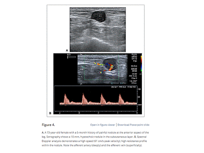
EXTRADIGITAL GLOMUS TUMOR


SWE of ROTATOR CUFF TENDON