Tổng số lượt xem trang

Thứ Bảy, 27 tháng 10, 2012

COLONIC POLYPS: US DETECTION


Colorectal cancer is often preventable if the precursor adenoma is detected and removed. Although ultrasound is clearly not one of the widely accepted screening techniques, this non-invasive and radiation-free modality is also capable of detecting colonic polyps, both benign and malignant. Such colon lesions may be encountered when not expected, usually during general abdominal sonography. The discovery of large colonic polyps is important and can potentially help reduce the incidence of a common cancer, whereas detection of a malignant polyp at an early stage may result in a curative intervention. This pictorial review highlights our experience of sonographic detection of colonic polyps in 43 adult patients encountered at our institutions over a 2-year period. 4 out of 50 discovered polyps were found to be malignant lesions, 3 polyps were hyperplastic, 1 polyp was a hamartomatous polyp and the rest were benign adenomas. The smallest of the detected polyps was 1.3 cm in diameter, the largest one was 4.0 cm (mean 1.7 cm; median 1.6 cm). In each case, polyps were discovered during a routine abdominal or pelvic examination, particularly when scanning was supplemented by a brief focused sonographic inspection of the colon with a 6–10 MHz linear transducer.


In this paper, we illustrate the key sonographic features of different types of commonly encountered colonic polyps in the hope of encouraging more observers to detect these lesions, which may be subtle.


Colonic polyps: sonographic morphology


Direct display of colonic polyps relies on demonstration of a spherical or ovoid hypoechoic lesion arising within colonic lumen (Figure 4). The hallmark for sonographic identification, in our experience, is the presence of demonstrable vascularity within such a lesion on Doppler.

When visible, colonic polyps usually lend themselves well to evaluation. On close inspection with a 6–10 MHz linear array transducer, surprisingly detailed views of the polypoid lesions can be achieved (Figure 5). The polyp pedicle is visualised as a prolongation of the mucosa, with a submucosal muscularis layer connecting the head of the polyp to the colonic wall (Figure 5d). Feeding vessels may also be seen. Adenomatous polyps may have smooth, or slightly convoluted, or lobular surface contour.
Even when the colonic lumen contains some echogenic residue, the polyps may be conspicuous owing to their low reflectivity (Figure 6). Echogenic faecal residue may also be moved away from the polyp by gentle compression with the transducer, which will improve visualisation.

Sessile polyps may be recognised when the lesion lies closely related to the colonic wall. Differentiation between pedunculated, sessile and flat polyps, however, is difficult unless the vascular stalk is readily visualised. When vascularity within the lesion is uncertain on inspection with colour Doppler, spectral Doppler analysis can confirm the presence of true vasculature (Figure 7). When no vascularity within the lesion is identified, then constant position, shape and size of such a lesion throughout the examination may suggest a polyp (Figure 8). Clearly, the observed lesion is unlikely to represent a polyp if it is avascular on Doppler and has moved with the transducer compression or in the course of the examination.
Discovery of a colonic polyp in a young individual will usually call for a targeted work-up for a polyposis syndrome. Hamartomatous polyps, classically associated with Peutz–Jeghers polyposis syndrome, may occasionally occur as isolated lesions (Figure 9).

Inflammatory polyps represent areas of elevated inflamed or normal mucosa. These may be sessile or pedunculated and are usually seen in patients with inflammatory disorders of the colon such as ulcerative colitis, Crohn's disease and dysenteric colitis. The term “pseudopolyp” is sometimes used to emphasise the non-neoplastic aetiology of these lesions [15]. Inflammatory polyps are similar to mucosa in echotexture, and thus recognised as extensions of the mucosa (Figure 10). Other changes indicating inflammatory bowel disease are usually present aiding differentiation.

Lipomas are also seen as intraluminal polypoid lesion, but can be recognised on ultrasound owing to their characteristic echogenic appearance.

The presence of malignancy may not be possible to estimate on ultrasound due to lack of reliable features. However, an apparent loss of wall stratification at the base of a large sessile polypoid lesion can suggest a carcinoma (Figure 11). The presence of a gas pocket retained within a large polyp indicates ulceration and is usually seen in malignant lesions (Figure 12). Detection of these features may warrant further CT investigation in addition to flexible sigmoidoscopy or colonoscopy.
 

Potential pitfalls


Several pitfalls that may lead to false-positive findings should be kept in mind when imaging the colon with ultrasound. These, in our experience, may be related to several factors, including a deficient scanning technique (such as tangential imaging), peculiar appearance of the colon owing to prominent convergent and bulbous haustral folds, presence of undigested food pieces and foreign bodies, and impacted diverticulum.

Tangential ultrasound imaging of the colonic wall in transverse plane, particularly in the areas of haustral folds, often results in false polypoid projections, which may be confusing (Figure 13a,b). This pitfall is avoided by multiplanar scanning of the colon with liberal transducer angulations that allow obtaining three-dimensional information.

Care should be taken not to confuse bulbous and complex haustral folds with polyps when the colon is imaged in longitudinal plane (Figure 13c). The linear, elongated nature of folds will aid differentiation in that circumstance. Another pitfall is to visualise a focal bulge in the colonic wall in cross-section that is present owing to prominent taeniae coli imaged during their contraction (Figure 14a).

Fragments of undigested food, pills or capsules and other foreign bodies may be occasionally encountered in the colon, but these are identified owing to their mobility, lack of vasculature and peculiar geometry, incompatible with that of polyps. The use of graded compression whereby the anterior and posterior walls of the bowel are opposed is helpful in displacing faeces and foreign bodies. A polyp may be reliably differentiated from faeces by the presence of continuation of mucosa and echogenic submucosa connecting the head of the polyp to the colonic wall (Figure 5d). The presence of a vascularised pedicle is also confirmatory of a polyp. In addition tiny cysts may be seen in the head of the polyp corresponding histologically to glands containing mucus [7].

Impacted diverticulae can be a potential source of confusion when they bulge prominently into the colonic lumen, but are readily recognised on ultrasound owing to the presence of trapped gas and inspissated stool (Figure 14b).

Fortunately, pitfalls leading to false-positives are usually avoided with careful technique. We did not encounter any false positive studies, which is in line with other observers reporting very high specificity of 99.4% for colonic polyp detection [7,8]. Of course, low sensitivity for detection of colonic polyps is a weakness of conventional ultrasound. Perhaps future advances in ultrasound imaging may someday permit this radiation-free non-invasive modality to play a much greater role in this area of colorectal imaging.

Conclusion


When integrated into routine scanning, brief sonographic examination of the accessible colon can reveal unsuspected large colonic polyps, which appear as spherical or ovoid, well-defined hypoechoic lesions within colonic lumen. Demonstration of vascularity within such lesions on Doppler is confirmatory. This may maximise the usefulness of conventional ultrasound and potentially help reduce the incidence of a common cancer since colonic polyps may harbour an early carcinoma or lead to malignancy.


Thứ Sáu, 26 tháng 10, 2012

HEPATORENAL INDEX in SCREENING for STEATOSIS


Abstract
OBJECTIVE. The hepatorenal index has been reported to be a sensitive and noninvasive test to quantify steatosis, but it is cumbersome and time-consuming and requires specialized software. The aim of this study was to improve and simplify the hepatorenal index calculation and determine whether it is an effective tool for differentiating patients with steatosis from those without steatosis, thereby eliminating the need for biopsy in a large number of patients.
MATERIALS AND METHODS. One hundred one patients who had undergone ultrasound-guided percutaneous liver biopsy at our institution were selected from a patient database. Patients with renal disease, patients with liver masses, and patients whose liver and right kidney were not included on the same image were excluded. Images were acquired with high-resolution ultrasound, and the hepatorenal index was calculated using freeware based on comparison of hepatic and renal brightness. 

RESULTS. Of the 101 patients, 63 had 5% or less steatosis and 38 had more than 5% steatosis. Using freeware available online from the National Institutes of Health, we calculated hepatorenal index values for all patients. Our data showed a strong correlation between the hepatorenal index and percentage of fat (r = 0.71, p < 0.0001). A hepatorenal index of 1.28 or greater had a 100% sensitivity for identifying more than 5% fat, 54% specificity, 0.57 positive predictive value, and 1.0 negative predictive value. If this method had been used prospectively to select patients for biopsy in our sample, 34% of biopsies could have been avoided. 

CONCLUSION. The hepatorenal index is a simple, reliable, and cost-effective screening tool for identifying patients who should not undergo liver biopsy for evaluation of steatosis.

FEMALE HYDROCELE [CYST of the CANAL of NUCK]

Nang ống Nuck


Discussion

Female hydrocele, also named a cyst of the canal of Nuck, is an unusual diagnosis with only about 400 reported cases. Because this entity is not
mentioned even in comprehensive surgical and gynecologic textbooks, many physicians are unaware of its very existence.

In the male fetus, an evagination of the parietal peritoneum, the processus vaginalis, accompanies the testis as it descends into the scrotum.

The same gloved fingerlike processus vaginalis, named the canal of Nuck after a 17th century Dutch anatomist, follows the round ligament of the uterus as it passes through the female inguinal canal. Normally this peritoneal evagination undergoes obliteration soon after birth in both sexes. If it remains completely patent, it forms an avenue for an indirect inguinal hernia.

Partial proximal obliteration, which leaves the distal portion of the processus vaginalis open, creates the anatomic prerequisite for a hydrocele of the spermatic cord and a cyst of the canal of Nuck, respectively. An imbalance of fluid secretion and absorption by the secretory membrane lining the processus vaginalis may eventuate in the development of a cystic swelling.

Hypersecretion or underabsorption may result from inflammation, trauma, or impairment of lymphatic drainage, for instance, owing to a large hernia, but in most cases it is idiopathic. The histologic composition of a cyst of the canal of Nuck reflects its peritoneal origin: a variously thick fibrous outer wall containing blood vessels and occasionally smooth muscle fibers, lined on the inside with a single layer of mesothelial cells. The diagnosis of a female hydrocele is seldom made on the basis of clinical findings alone. In fact, most of the cases reported in the literature were diagnosed at surgery performed for suspected inguinal hernias. Patients usually have a painless or moderately painful fluctuant inguinal mass, which is irreducible and can be transilluminated if large enough. There are no acute abdominal symptoms as with an incarcerated inguinal hernia, for which it is often mistaken. Clinical diagnosis is even made more difficult by the fact that in one third of the patients, a concomitant inguinal hernia is found. There are many differential diagnoses for groin masses. Most commonly they are caused by inguinal or femoral hernias, followed by enlarged lymph nodes and soft tissue tumors (eg, lipomas, leiomyomas, and endometriosis of the round ligament). Further down the list are vascular abnormalities (eg, arterial and venous aneurysms) and some very rare entities such as ganglion cysts protruding out of the hip joint and paraspinal abscesses surfacing in the groin.  High-resolution sonography can identify the nature of groin tumors in most cases or at least can narrow down the list of differential diagnoses.

To the best of our knowledge, there has been only 1 report dealing with the sonographic appearance of hydrocele of the canal of Nuck.

Anderson et al  found a tubular anechoic mass extending along the course of the round ligament without any internal structures, thus representing a unilocular hydrocele. Miklos et al described sonographically guided placement of a hook-wire needle to facilitate surgical exposure of a multicystic hydrocele of the canal of Nuck without further detailing its sonographic features.

Multiloculated hydroceles are reported in female as well as in male patients, so internal septations are obviously not uncommon. As for the somewhat strange internal structures in our cases (cysts within cysts), we can only speculate. We assume that they represent remnants of a futile attempt by nature at obliteration.

There are few conditions that can be confused with hydrocele of the canal of Nuck on sonography. Malignant lymphomas may at first glance have the appearance of anechoic cysts but show abundant vasculature on color-coded Doppler sonography. Generally, we would suggest color Doppler interrogation of all seemingly cystic structures to rule out a vascularized structure before performing interventional measures. An incarcerated hernia contains fluid, but there is always a solid, mostly hyperechoic component protruding out of the hernial orifice into the sac, representing compressed omentum or intestine (Figure 3).
Abscesses, apart from occurring in an entirely different clinical setting, usually have an irregular hypoechoic wall and echogenic debris or gas bubbles in their contents. The peculiar sonographic morphologic features of hydrocele of the canal of Nuck, namely, a cystic mass with a fine circumferential echogenic margin, possibly with septa or cystic internal structures, make high-resolution sonography the imaging modality of choice for diagnosing this entity.

In conclusion, hydrocele of the canal of Nuck is a rare developmental disorder, but it ought to be on the differential diagnosis list of groin tumors in female patients. The ultrasound community especially should be aware of this entity because this diagnosis can readily and confidently be established on the basis of sonography. A concomitant inguinal hernia can be shown or ruled out in the same way. Although surgical excision is curative and therefore the treatment of choice, in our opinion, sonographically guided aspiration can be used to temporarily alleviate patient discomfort in this absolutely benign condition.

Thứ Năm, 25 tháng 10, 2012

MIRROR-IMAGE ARTIFACT on TVS


A 27-year-old woman was admitted to the obstetric emergency department for abdominal pain without bleeding at 8 weeks’ gestation. She had a previous uneventful pregnancy delivered vaginally at term, and an early scan performed 2 weeks before the symptoms occurred revealed an ongoing intrauterine pregnancy.

The patient underwent transvaginal sonography (MyLab 25; Esaote SpA, Florence, Italy), which showed an intrauterine pregnancy with a live embryo measuring 29 mm, consistent with a gestational age of 8 weeks 5 days. A second gestational sac with irregular and undefined boundaries containing an active embryo measuring 17 mm was depicted in the rectouterine pouch (Figure 1). Both ovaries were visualized as normal, and no pelvic free fluid collection was noticed during the scan. These findings were consistent with a diagnosis of heterotopic pregnancy.

Heterotopic pregnancy refers to the rare occurrence of both intrauterine and ectopic pregnancies usually located in one fallopian tube, cervix, or, more rarely, abdomen. Assisted reproduction techniques, tubal surgery, pelvic inflammatory disease, and the use of intrauterine devices represent the most common risk factors.1

Sonography is the mainstay for diagnosis of heterotopic pregnancy, allowing for the detection of two gestational sacs located inside and outside the uterus, respectively, and blood collection in the pelvis. In addition, clinical symptoms such as pain and genital tract bleeding can help in achieving the diagnosis.1

In this case, the patient had no risk factors for this condition, with a previous uneventful pregnancy delivered at term and no history of pelvic surgery or disease. To confirm the diagnosis, the woman was asked to fill her bladder, and transabdominal sonography was performed. Interestingly, only the intrauterine gestational sac was found, with a normal appearance of the rectouterine pouch and no ectopic pregnancy detected.

AVID


Abstract

A series of 20 cases from 2 academic institutions is presented with a characteristic imaging triad of asymmetric ventriculomegaly, a large interhemispheric cyst, and partial or complete agenesis of the corpus callosum. Most cases were initially referred as aqueduct stenosis and hydrocephalus or focal porencephaly. We describe the imaging findings that identify an abnormal or absent corpus callosum associated with a type 1 interhemispheric cyst in fetuses initially thought to have hydrocephalus attributable to aqueductal stenosis. We suggest that the acronym AVID (asymmetric ventriculomegaly, interhemispheric cyst, and dysgenesis of the corpus callosum) may be useful in recognition of these cases. All cases presented with markedly asymmetric ventriculomegaly on initial sonography, with progressive hydrocephalus throughout gestation. Fetal magnetic resonance imaging was performed in 15 of 20 cases. Thirteen of 20 cases were identified in male fetuses. Associated fetal and postnatal abnormalities are also reported. Technological improvements in sonography and fetal magnetic resonance imaging allow improved characterization of associated intracranial anomalies in the setting of hydrocephalus. Accurate diagnosis can aid parental counseling, especially because isolated aqueductal stenosis suggests a better prognosis than hydrocephalus with anomalies. Markedly asymmetric ventriculomegaly in this series was the key to excluding isolated aqueductal stenosis and was associated with callosal malformation with a type 1a interhemispheric cyst.


Thứ Tư, 24 tháng 10, 2012

EVALUATION and MANAGEMENT of TIPS


Abstract

OBJECTIVE. The purpose of this article is to describe the evaluation of transjugular intrahepatic portosystemic shunts (TIPS) and the management of dysfunctional shunts.


CONCLUSION. TIPS can become dysfunctional if stenosis develops in the shunt or the hepatic vein above the shunt. Screening allows detection of stenoses before portal hypertensive–related complications recur. Revision of stenotic shunts can be easily accomplished in most cases. Techniques for screening and revision will be discussed.

Although transjugular intrahepatic portosystemic shunts (TIPS) have become integral to the management of portal hypertensive–related complications, stenosis of the shunt has been a major problem. Originally, when TIPS were all created with bare metal stents, the loss of primary patency was around 50% at 1 year after shunt creation. The introduction of polytetrafluoroethylene (PTFE)–covered stents has vastly improved patency but stenosis still occurs in 8–20% of patients at 1 year after TIPS creation [15].

The significance of TIPS stenoses is that they can lead to recurrent portal hypertension and put patients at risk for reaccumulation of ascites or further variceal hemorrhage. It is important for interventional radiologists to actively follow their patients with TIPS to assess how well the shunt is functioning. The interventional radiologists understand the shunt better and should be able to make better assessments regarding the function of the TIPS and the need for revision. Furthermore, if you place the shunt and have others follow the patient, then your role as physician is diminished to that of technician.

Evaluating TIPS Function

There is no universal agreement on protocol for when or how often to screen TIPS function. One approach is to simply wait for symptoms of portal hypertension to recur. This approach can be used in patients whose TIPS were placed to treat ascites. This approach is not advisable for patients with a history of variceal bleeding because the first symptom suggesting a problem with the TIPS might be a fatal recurrent hemorrhage. Even in patients with ascites, detecting shunt problems before the patient becomes especially symptomatic is beneficial because TIPS venography is much easier when the patient does not have a large volume of ascites. The patient is more comfortable and able to breathe easier in a supine position when there is not much ascites. Furthermore, massive ascites forces the liver more cephalad, and the extra density caused by the ascites degrades the fluoroscopic image; both of these effects make the procedure more difficult.

The timing of screening has also not been standardized and varies greatly between institutions. Our protocol has been to perform Doppler ultrasound at 1, 3, 6, and 12 months after TIPS creation and every 6–12 months thereafter, depending on the patient’s clinical status. Evaluations at other time intervals may be triggered by any recurrence of ascites or bleeding. Although this schedule has not been scientifically validated, it has served us well in our relatively large TIPS experience (1223 patients since 1991) and is similar to protocols used by others. Through use of this protocol, many hemodynamically significant TIPS stenoses have been detected and fixed before the patients experienced recurrent hemorrhage. In recent years, the improved patency of TIPS made possible by the development of a dedicated PTFE-covered device (Viatorr, W. L. Gore) has allowed some loosening of this schedule and even caused some authors [6] to speculate that routine screening may not be necessary for TIPS created with Viatorr stents. Again, the ideal timing of follow-up screening in this era of stent-grafts has not been scientifically determined.

Ultrasound has been the primary tool used to screen for TIPS stenoses because it is noninvasive, readily available, and relatively low cost compared with other imaging modalities. However, many different ultrasound parameters have been used to assess patency, with variable results.

Flow velocities in the TIPS represent the primary parameter. A single velocity measurement in the mid shunt has been used by some, although the sensitivity and specificity with this methodology were only 86% and 54%, respectively, in one study [7]. However, with careful examination of velocities along the length of the shunt, it is often possible to identify a specific stenosis with a major change in velocities across the stenosis. Whereas some authors [79] use a drop in shunt velocities to below 40–60 cm/s as the criterion for calling a stenosis, it is also possible to measure significantly elevated velocities (over 200 cm/s) in a jet-effect zone just beyond the stenosis (Figs. 1A, 1B, 1C, 1D, 1E, 1F, 1G, and 1H).
 
 
However, if the stenosis is very close to the inferior vena cava (IVC) within the hepatic vein, the jet effect will not be seen and the velocities will be uniformly low within the TIPS (Figs. 2A, 2B, 2C, and 2D). Thus, velocities in the shunt should range between 90 and 190 cm/s in most patent TIPS, and peak velocities below or above this range may indicate a stenosis [10].


Main portal vein velocity is another useful parameter. Our group previously showed that before TIPS the main portal velocity is usually 20 cm/s but after TIPS it typically increases to more than 30 cm/s [11]. When a shunt gets stenotic, the flow in the portal vein leading up to the TIPS is diminished and the main portal velocity drops often down below 30 cm/s. Others have used higher values, such as 40 cm/s [12], but they have also reported ultrasound to be an inaccurate screening tool. The direction of flow in the portal vein branches should also be evaluated. In most patients who have pre-TIPS hepatopetal flow (toward the liver), the flow direction reverses and becomes hepatofugal after creation of the TIPS. When a stenosis develops, the flow in these branches often reverts to hepatofugal [13].

We have routinely used ultrasound with a high degree of confidence to screen TIPS function. In a study that compared ultrasound criteria to venographic proof of stenosis, it was found that no individual parameter was more than 84% specific in predicting TIPS dysfunction. However, when an overall assessment was made by considering all the parameters, the sensitivity and specificity for detecting TIPS stenoses were 92% and 72%, respectively [10]. Also in that study, it was shown that when both main portal velocity and distal shunt velocity are abnormal, ultrasound has 100% specificity for detection of TIPS malfunction. Furthermore, it is important to follow these numbers over time because initially after TIPS, the velocities may all be normal, but changes in velocities and flow directions can indicate that a stenosis has developed. In fact, Dodd et al. [14] considered temporal changes in velocity more sensitive than static low-velocity parameters. Other investigators [13, 1518] have supported that ultrasound is very sensitive for detecting shunt malfunction.

Unfortunately, not all investigators have found ultrasound to be especially useful. One prospective double-blinded study reported that ultrasound predicted shunt patency in 20 of 31 shunts that proved to be occluded or stenotic [9]. However, their main criterion for calling a stenosis was a peak shunt velocity of < 60 cm/s, which is somewhat simplistic. Using more extensive velocity criteria, another study [12] still found concordance between ultrasound and venography in only 53% of cases, and in their experience ultrasound rarely predicted a stenosis that was not already suspected on clinical grounds. Given the variability in the reported sensitivity and specificity of ultrasound, it is important to evaluate the results at your own institution to see how your ultrasound readings correlate with venography and pressure measurements.

The use of echo enhancers has been proposed as a way of improving the accuracy of ultrasound. In a small study of 31 TIPS, the use of echo enhancers was found to increase the specificity of ultrasound from 89% to 100% [19]. However, this technique has not become common practice.


Thứ Bảy, 20 tháng 10, 2012

IBD in Pediatric Patient

 
Background and Importance

IBD is one of the most common gastrointestinal diseases affecting pediatric patients in the developed world [1]. Crohn disease (CD) and ulcerative colitis (UC) are the two predominant subtypes of IBD, differing both in distribution of gastrointestinal tract involvement and depth of inflammation. Both disorders are most common in Europe and North America, where the ranges of incidence and prevalence are 3.1–14.6 cases per person-years and 26–199 cases per 100,000 persons for CD and 2.2–14.3 cases per 100,000 person-years and 37–246 cases per 100,000 persons for UC [1]. The classic teaching is that CD has a bimodal peak—the first peak in the second or third decade of life and a smaller second peak in the sixth or seventh decade. There is equal evidence, however, of a unimodal peak in the second or third decade that explains the high incidence in the adolescent population [1, 2]. CD is more common than UC among adolescents, and adolescents with UC tend to have more severe and extensive disease at presentation than their adult counterparts do: Approximately 90% of adolescents present with total colonic involvement [3]. Because the bowel disease in UC is confined to the colon, total colonic surgical resection is curative for patients whose condition is refractory to medical therapy. In contrast, the potential involvement of the entire gastrointestinal tract in CD often leads to lifelong intermittent symptomatic recurrence, and medical rather than surgical management is the primary therapy. No consensus exists regarding the optimal technique and imaging modality for evaluating IBD. The choice of imaging is informed by the clinical presentation of the patient. The choice of specific modality is based on the need to assess the distribution or activity of the disease and to detect extraluminal complications, such as intraabdominal abscess, perforation of bowel, and enteric fistula. Pediatric patients need additional attention to minimization of radiation exposure during imaging examinations because the chronic remitting and relapsing nature of IBD, especially CD, frequently necessitates repeat imaging with a resultant greater cumulative lifetime radiation exposure.


Ultrasound has the advantage of being a noninvasive test that imparts no ionizing radiation. High interoperator variability, however, is a practical consideration for determining its true diagnostic accuracy.

Targeted assessment of the bowel wall is usually performed with a high-frequency linear-array probe. As with fluoroscopy, with ultrasound, bowel loops can be observed over time for evaluation of peristalsis and function.
 

The ultrasound criteria for assessment of CD include assessment of wall thickness, loop stiffness, bowel dilatation, presence or absence of strictures, abnormal peristalsis, presence of fistula or abscess, and mesenteric inflammatory change. A previous meta-analysis revealed mean per patient sensitivity of 89.7% and specificity of 95.6% and per bowel segment sensitivity and specificity of 73.5% and 92.9%. Patient preparation is not usually required, although the studies are usually performed after the patient has fasted. Use of contrast material has been found to increase accuracy, and Doppler evaluation of bowel wall vascularity may help to determine the presence of disease activity or quiescence.


Poor visualization of the rectum and sigmoid colon, owing to the location of these structures in the pelvis, makes ultrasound less suitable for assessment of UC. The spatial resolution of ultrasound also is lower than that of barium studies.


In all of the ultrasound studies, wall thickness was the principal imaging parameter. The sensitivities and specificities were consistently much lower than with the cross-sectional modalities; the range of sensitivity was 0.48–0.8 and that of specificity was 0.57–0.93 across the three papers.

Ultrasound yields useful information on bowel wall abnormalities and can be accurate in experienced hands; however, because of low sensitivity and specificity and high interoperator variability, ultrasound is not recommended for first-line imaging in the three clinical scenarios posed.

The Three Typical Clinical Scenarios

1/ Pediatric patient: initial diagnosis of suspected inflammatory bowel disease, differentiation of Crohn disease from ulcerative colitis.

2/ Pediatric patient: known inflammatory bowel disease with new acute presentation (fever, peritonitis, leukocytosis).

3/ Pediatric patient: known inflammatory bowel disease with symptomatic recurrence (abdominal pain, diarrhea), not acutely ill.

Xem SIÊU ÂM TRONG BỆNH LÝ VIÊM RUỘT (IBD)


Thứ Ba, 16 tháng 10, 2012

FIRST US TOOL for DENSE BREAST


Clinical Context

The National Cancer Institute estimates that approximately 40% of women undergoing screening mammography have dense breast tissue. High breast density hinders mammographic interpretation, which may delay detection of breast cancer until it is at a more advanced and difficult-to-treat stage.
Less-dense breasts have a high amount of fatty tissue, whereas dense breasts have a high amount of connective and fibroglandular tissue. Breast cancer and fibroglandular breast tissue both appear as solid white areas on mammograms, rendering interpretation more difficult.

Study Synopsis and Perspective

The US Food and Drug Administration (FDA) approved the first ultrasound device for use in combination with mammography in women with dense breast tissue.
The device, known as the somo-v Automated Breast Ultrasound System (ABUS), provides clinicians with an additional resource in screening women with dense breasts. The indication is limited to use in women who have a negative mammogram result and no symptoms of breast cancer.
Mammograms of dense breasts can be difficult to interpret, the FDA points out in its announcement of the approval.
"A physician may recommend additional screening using ultrasound, for women with dense breast tissue and a negative mammogram," said Alberto Gutierrez, PhD, director of the Office of In Vitro Diagnostic Device Evaluation and Safety at the FDA's Center for Devices and Radiological Health. "The somo-v ABUS is a safe and effective breast ultrasound tool when such screening is recommended."
Approximately 40% of women undergoing screening mammography have dense breasts, according to National Cancer Institute estimates. These women have an increased risk for breast cancer, with detection usually at a more advanced and difficult-to-treat stage, the FDA said.
In April, an expert advisory committee of the FDA voted unanimously to recommend the expanded use of the ultrasound device as a screening tool for women with dense breast tissue.
However, as reported by Medscape Medical News, some of the panel members had concerns about the device because of its automated nature. In contrast to handheld ultrasound, medical specialists who operate an automated ultrasound do not have to be imaging experts, Robert Faulk, MD, from Medical Imaging Consultants in Omaha, Nebraska, noted during the meeting. "For instance, obstetricians and gynecologists could do ABUS and interpret the results themselves, along with mammography findings," Dr. Faulk said. "That could have a deleterious effect on healthcare for many women," he added.
"ABUS as a screening tool could potentially be applied to 40 million women in the United States [with dense breasts], and if it were used by nonimaging specialists, the false-positive rates could go through the ceiling," said Daniel Kopans, MD, from Harvard Medical School in Boston, Massachusetts. Dr. Kopans commented that postmarket surveys of the false-positive rate from the new ultrasound device could provide important data on the effect of its use as a screening tool in women with dense breasts.
Dense breasts have a high amount of connective and fibroglandular tissue compared with less-dense breasts, which have a high amount of fatty tissue, the FDA explained.
Fibroglandular breast tissue and tumors both appear as solid white areas on mammograms, which can complicate interpretation. Dense breast tissue may obscure smaller tumors, potentially delaying detection of breast cancer, according to the FDA.
Ultrasound imaging has been proven capable of detecting small masses in dense breasts.
A clinical study has shown a statistically significant increase in breast cancer detection when images generated by this new device were reviewed in conjunction with mammograms compared with mammograms alone. The study involved board-certified radiologists who reviewed mammograms alone or in conjunction with device-generated images for 200 women with dense breasts and negative mammograms.
As part of the approval, the FDA requires that the manufacturer train clinicians and technologists using the new ultrasound device, and that the manufacturer provide each facility with a manual clearly defining system tests required for initial, periodic, and yearly quality-control measures.
The ultrasound works via a transducer that directs high-frequency sound waves at the breast. The specially shaped transducer of the device can automatically scan the entire breast in approximately 1 minute to produce several images for review, according to the FDA press materials.
More information on the new ultrasound device is available on the FDA Web site.


Clinical Implications

  • The FDA has approved a new automated ultrasound device to be used in combination with mammography in women with dense breast tissue. This is the first ultrasound device approved for this indication. The specially shaped transducer of the device directs high-frequency sound waves at the breast to scan the entire breast in approximately 1 minute, automatically producing several images for review.
  • The FDA has approved the ultrasound device for use only in women with negative mammography results and no symptoms of breast cancer. In these women, the device is a safe and effective breast ultrasound tool. Compared with mammograms alone, mammograms plus images generated by the new device interpreted by board-certified radiologists were associated with a statistically significant increase in breast cancer detection.
  • Unlike handheld ultrasound, automated ultrasound does not have to be operated by an imaging expert. This may be a potential drawback in that medical practitioners without specialized radiographic training could use the new device and interpret the results themselves, along with mammography findings. This could dramatically increase false-positive rates. The FDA is requiring that the manufacturer train clinicians and technologists using the new ultrasound device, and that each facility is given a manual clearly defining system tests required for quality control.


    Abstract: The idea of an automated whole breast ultrasound was developed three decades ago. We present our initial experiences with the latest technical advance in this technique, the automated breast volume scanner (ABVS) ACUSON S2000TM. Volume data sets were collected from 50 patients and a database containing 23 women with no detectable lesions in conventional ultrasound (BI-RADS®-US 1), 13 women with clearly benign lesions (BI-RADS®-US 2), and 14 women with known breast cancer (BI-RADS®-US 5) was created. An independent examiner evaluated the ABVS data on a separate workstation without any prior knowledge of the patients’ histories. The diagnostic accuracy for the experimental ABVS was 66.0% (95% confidence interval [CI]: 52.9–79.1). The independent examiner detected all breast cancers in the volume data resulting in a calculated sensitivity of 100% in the described setting (95% CI: 73.2%–100%). After the ABVS examination, there were a high number of requests for second-look ultrasounds in 47% (95% CI: 30.9–63.5) of the healthy women (with either a clearly benign lesion or no breast lesions at all in conventional handheld ultrasound). Therefore, the specificity remained at 52.8% (95% CI: 35.7–69.2). When comparing the concordance of the ABVS with the gold standard (conventional handheld ultrasound), Cohen’s Kappa value as an estimation of the inter-rater reliability was κ = 0.37, indicating fair agreement. In conclusion, the ABVS must still be regarded as an experimental technique for breast ultrasound, which definitely needs to undergo further evaluation studies.

    Keywords: breast cancer, automated breast ultrasound, automated breast volume scanner, ABVS

Chủ Nhật, 14 tháng 10, 2012

NHÂN CA OVARIAN TERATOMA tại MEDIC


Figure 4d.  Mature cystic teratoma of the right ovary in a 19-year-old pregnant woman. (d) Photograph of the gross specimen shows yellowish, pasty sebaceous material (black arrowhead) and hair (white arrowheads) within the cyst cavity, findings that account for the fat echogenicity and signal intensity seen at US and MR imaging. Two molar teeth are also evident (arrows).
 
Figure 4(a) Sagittal transabdominal US image shows an echogenic mass with sound attenuation (arrows). 
 
Abstract
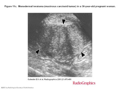
Ovarian teratomas include mature cystic teratomas (dermoid cysts), immature teratomas, and monodermal teratomas (eg, struma ovarii, carcinoid tumors, neural tumors). Most mature cystic teratomas can be diagnosed at ultrasonography (US) but may have a variety of appearances, characterized by echogenic sebaceous material and calcification.


At computed tomography (CT), fat attenuation within a cyst is diagnostic. At magnetic resonance (MR) imaging, the sebaceous component is specifically identified with fat-saturation techniques. The US appearances of immature teratoma are nonspecific, although the tumors are typically heterogeneous, partially solid lesions, usually with scattered calcifications.


At CT and MR imaging, immature teratomas characteristically have a large, irregular solid component containing coarse calcifications. Small foci of fat help identify these tumors. The US features of struma ovarii are also nonspecific, but a heterogeneous, predominantly solid mass may be seen. On T1- and T2-weighted images, the cystic spaces demonstrate both high and low signal intensity. Familiarity with the US, CT, and MR imaging features of ovarian teratomas can aid in differentiation and diagnosis.
.....
Most mature cystic teratomas can be diagnosed at US. However, the US diagnosis is complicated by the fact that these tumors may have a variety of appearances. Three manifestations occur most commonly. The most common manifestation is a cystic lesion with a densely echogenic tubercle (Rokitansky nodule) projecting into the cyst lumen (16). The second manifestation is a diffusely or partially echogenic mass with the echogenic area usually demonstrating sound attenuation owing to sebaceous material and hair within the cyst cavity (Fig 4) (17),(18). The third manifestation consists of multiple thin, echogenic bands caused by hair in the cyst cavity (Fig 3). Pure sebum within the cyst may be hypoechoic or anechoic (19). Fluid-fluid levels result from sebum floating above aqueous fluid, which appears more echogenic than the sebum layer (18). The dermoid plug is echogenic, with shadowing due to adipose tissue or calcifications within the plug or to hair arising from it. Diffuse echogenicity in these tumors is caused by hair mixed with the cyst fluid (Fig 4).
 
In a prospective US study that made use of these criteria, Mais et al (20) found a sensitivity of 58% and a specificity of 99% in the diagnosis of mature cystic teratoma. Numerous pitfalls have been described in the US diagnosis of mature cystic teratoma (21). Blood clot within a hemorrhagic cyst can appear echogenic, although a mature cystic teratoma usually demonstrates sound attenuation rather than increased through-transmission. Hemorrhagic cysts or blood clots typically demonstrate increased through-transmission. Echogenic bowel can frequently be mistaken for diffusely echogenic mature cystic teratoma and vice versa (21). Perforated appendix with appendicolith and fibrous lesions such as cystadenofibromas have also been described as false-positive findings (21),(22).
 © RSNA, 2001