
Purpose: Objective Shear wave elastography (SWE) enabled living tissue assessment of stiffness. This is routinely used for breast, thyroid and liver diseases, but there is currently no data for the brain. We aim to characterize elasticity of normal brain parenchyma and brain tumors using SWE.
Materials and Methods: Patients with scheduled brain tumor removal were included in this study. In addition to standard ultrasonography, intraoperative SWE using an ultrafast ultrasonic device was used to measure the elasticity of each tumor and its surrounding normal brain. Data were collected by an investigator blinded to the diagnosis. Descriptive statistics, box plot analysis as well as intraoperator and interoperator reproducibility analysis were also performed.
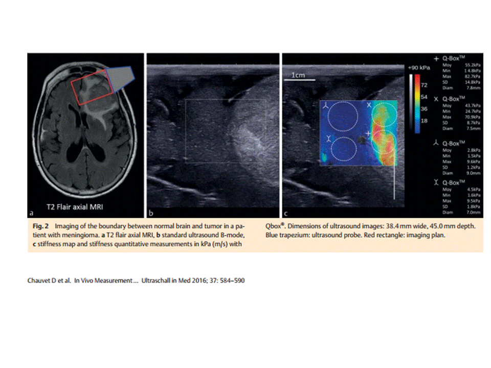
Results: 63 patients were included and classified into four main types of tumor: meningiomas, low-grade gliomas, high-grade gliomas and metastasis. Young’s Modulus measured by SWE has given new insight to differentiate brain tumors: 33.1 ± 5.9 kPa, 23.7 ± 4.9 kPa, 11.4 ± 3.6 kPa and 16.7 ± 2.5 kPa, respectively, for the four subgroups. Normal brain tissue has been characterized by a reproducible mean stiffness of 7.3 ± 2.1 kPa. Moreover, low-grade glioma stiffness is different from high-grade glioma stiffness (p = 0.01) and normal brain stiffness is very different from low-grade gliomas stiffness (p under 0.01).
Conclusion: This study demonstrates that there are significant differences in elasticity among the most common types of brain tumors. With intraoperative SWE, neurosurgeons may have innovative information to predict diagnosis and guide their resection.


Không có nhận xét nào :
Đăng nhận xét