April 19, 2023
Researchers led by Dr. Leila Aghaghazvini from the Iran University of Medical Sciences in Tehran found that using these measures adds diagnostic value to traditional TI-RADS assessment of suspicious thyroid nodules, including higher sensitivity and specificity among other achievement marks.
"It would be advantageous to incorporate these criteria into TI-RADS risk stratification systems in order to reduce unnecessary fine-needle aspirations while keeping a high detection sensitivity for malignant tumors," Aghaghazvini and colleagues wrote.
Conventional ultrasound is typically used to detect thyroid cancer after physical examination and palpitation. However, Aghaghazvini et al noted that there is a lack of evidence that any particular ultrasound findings can predict thyroid cancer.
Specialists use TI-RADS to classify risk of malignancy based on ultrasound findings. But Aghaghazvini said that this does take a nodule's vascular or elastic characteristics into account. To address this knowledge gap, the team explored the value of adding vascularity and shear-wave elastography assessments to conventional TI-RADS assessments.
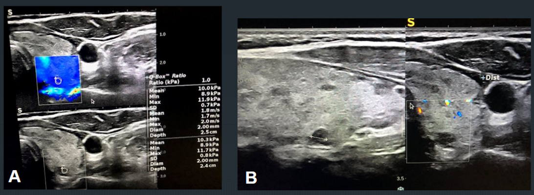
The study included 200 total thyroid nodules, assessed by conventional sonography to gather TI-RADS scores and important characteristics. The investigators used a 7.5 MHz probe to assess vascularity pattern and resistive index, performed quantitative elastography evaluations via color mapping, and calculated average and maximum velocities. They confirmed final diagnoses of all thyroid nodules via histopathology assessment or follow-up imaging.
The study found 37 TI-RADS 4 nodules, 15 of which were malignant, and 57 TI-RADS 5 nodules, 12 of which were malignant.
Overall, area under the curve (AUC) for TI-RADS assessment was 0.76. Adding elastography and resistive index measures to the mix translated to higher AUC values for TI-RADS 4 and 5 nodules.
| AUC values of vascularity and elastography measures | ||
| Measure | TI-RADS 4 | TI-RADS 5 |
| Resistive index | 0.93 | 0.96 |
| Color map elastography grades | 0.89 | 0.85 |
| Shear-wave elastography maximum velocity | 0.86 | 0.96 |
| Shear-wave elastography average velocity | 0.82 | 0.97 |
| Doppler grade | 0.8 | 0.8 |
The researchers also used a resistive index cutoff point of 0.6 in TI-RADS 4 nodules and found even higher values. This included AUCs of 0.93 for sensitivity, 1 for specificity, 0.97 for efficiency, 1 for positive predictive value, and 0.96 for negative predictive value.
When a cutoff point of 4.33 was used for shear-wave elastography average velocity, the researchers observed high diagnostic efficacy values. These included AUCs of 1 for sensitivity, 0.82 for specificity, 0.86 for efficiency, 0.6 for positive predictive value, and 1 for negative predictive value.
The study authors hope that these assessments will improve differentiation between malignant and benign thyroid nodules, Aghaghazvini said.
Không có nhận xét nào :
Đăng nhận xét