Tổng số lượt xem trang

Chủ Nhật, 18 tháng 2, 2018

Acute Flank Pain: Renal Stones and Acute Pyelonephritis

Abstract

Ultrasonography is a useful tool for the differential diagnosis of acute flank pain. Renal stones appear as a focal area of echogenicity with acoustic shadowing on ultrasonography. In acute pyelonephritis (APN), the kidneys may be enlarged and have a hypoechoic parenchyma with loss of the normal corticomedullary junction. However, clinical and laboratory correlations are essential for the diagnosis of renal stones and APN through imaging studies. This review describes the typical ultrasonography features of renal stones and APN. Moreover, in daily practice, cross-sectional imaging is essential and widely used to confirm renal stones and APN and to differentiate them from other diseases causing flank pain. Other diseases causing acute flank pain are also described in this review.


Thứ Sáu, 9 tháng 2, 2018

ROTATOR INTERVAL


Abstract

 Shoulder pathologies of the rotator cuff of the shoulder are common in clinical practice. The focus of this pictorial essay is to discuss the anatomical details of the rotator interval of the shoulder, correlate the anatomy with normal ultrasound images and present selected pathologies. We focus on the imaging of the rotator interval that is actually the anterosuperior aspect of the glenohumeral joint capsule that is reinforced externally by the coracohumeral ligament, internally by the superior glenohumeral ligament and capsular fibers which blend together and insert medially and laterally to the bicipital groove. In this article we demonstrate the capability of high-resolution musculoskeletal ultrasound to visualize the detailed anatomy of the rotator interval. MSUS has a higher spatial resolution than other imaging techniques and the ability to examine these structures dynamically and to utilize the probe for precise anatomic localization of the patient’s pain by sono-palpation.


Thứ Năm, 8 tháng 2, 2018

Zika's effects hard to detect on ultrasound, MRI


February 5, 2018 -- A new animal study published February 5 in Nature Medicineillustrates the challenges in detecting how the Zika virus affects the fetuses of infected mothers. Researchers found that the Zika virus caused injury to the brains of fetal macaque monkeys that was hard to detect with ultrasound and even MRI.

Zika sent chills throughout tropical regions when cases began appearing frequently in 2016, with Brazil being hit particularly hard. Most often spread by mosquitoes, the disease caused conditions such as microcephaly in infants born to infected mothers. While the outbreak has subsided since then, healthcare providers and public health authorities are keeping a watchful eye for its resurgence.
In the new paper, researchers from multiple centers described their efforts to detect the signs of Zika damage in the brains of fetuses by using tools including ultrasound and MRI (Nat Med, February 5, 2018). Knowledge of these signs could help providers counsel pregnant women with Zika infection, in particular for infants who are infected with the virus but who don't have the telltale signs of microcephaly.
The researchers infected a test group of five pregnant pigtail macaque monkeys, while a control group of animals remained uninfected. They followed the symptoms that emerged after infection and performed regular imaging studies with ultrasound and MRI, in addition to using blood tests and other exams to track the progress of the monkeys and their fetuses.
Weekly ultrasound scans detected no obvious fetal abnormalities except for a periventricular echogenic lesion and ventriculomegaly in one monkey, the group found. Likewise, Doppler ultrasound scans of the middle cerebral artery in the fetuses showed no differences in the resistance index, which the researchers took to mean that fetal brain oxygenation was the same between the infected and control monkeys.
One imaging tool that did work was MRI with a half-Fourier acquisition single-shot turbo spin-echo (HASTE) T2-weighted protocol. The technique revealed periventricular subcortical T2-hyperintense foci in the posterior brain in four of the five animals at 120 to 129 days of gestation, signs that were not found in the control fetuses. The brainstem and cerebellum otherwise appeared normal except for a cyst in one of the infected fetuses.
Signs of Zika on serial MRI scans
Researchers found telltale signs of Zika (periventricular subcortical T2-hyperintense foci in the posterior brain) on serial brain MRI scans with a HASTE protocol but said the phenomena were transient.
The study shows the difficulty in detecting signs of brain injury in fetuses carrying the Zika virus, the researchers concluded. While T2-hyperintense foci were detected by MRI, the phenomenon was transient, and in any event MRI is not widely available in many regions where Zika is prevalent.

"Our study highlights the inability of standard prenatal diagnostic tools to detect silent pathology in the fetal brain associated with congenital Zika infection in the absence of microcephaly," they wrote.

Thứ Bảy, 3 tháng 2, 2018

Low-risk thyroid nodules safe to track with ultrasound


By Kate Madden Yee, AuntMinnie.com staff writer
February 1, 2018 -- It's safe to use ultrasound to track thyroid nodules that don't have suspicious features after fine-needle aspiration (FNA) biopsy -- although nodules with intermediate or highly suspicious features should be evaluated with another biopsy, according to research in the February issue of the American Journal of Roentgenology.

The findings address disagreement among researchers regarding how to handle nondiagnostic nodules, defined as nodules that can't be classified as malignant or benign after FNA biopsy, wrote a team led by Dr. Chae Jung Park of Yonsei University College of Medicine in Seoul, South Korea. Some professional groups recommend repeat fine-needle aspiration biopsy if a nondiagnostic nodule is solid at ultrasound examination, while others say this isn't necessary.
"Some researchers suggest that clinical and ultrasound follow-up may be more appropriate than repeat fine-needle aspiration after nondiagnostic biopsy regardless of ultrasound features because few malignancies are diagnosed in this population," the study authors wrote. "The purposes of this study were to evaluate the malignancy rate of nodules with nondiagnostic cytologic results ... and to establish management guidelines for these nodules according to the ultrasound patterns."
Safe and reliable
Ultrasound-guided FNA biopsy is considered a safe, reliable, and cost-effective way to distinguish between benign and malignant thyroid nodules. But the procedure can produce a nondiagnostic result if the sample isn't adequate, the group wrote (AJR, February 2018, Vol. 210:2, pp. 412-417).
In 2015, the American Thyroid Association (ATA) recommended repeat FNA biopsy for initially nondiagnostic nodules and surgical confirmation for nodules with suspicious ultrasound features, along with continued close follow-up with ultrasound for repeatedly nondiagnostic nodules without suspicious ultrasound features.
However, these guidelines have not been confirmed, Park and colleagues wrote.
"To our knowledge, no validation studies have been conducted to assess these management guidelines according to the 2015 ATA ultrasound patterns," they wrote.
Park's group assessed 441 nodules larger than 1 cm found in 437 patients with nondiagnostic ultrasound results between January 2013 and December 2014. Of these 441 nodules, 191 were confirmed with cytopathology or were smaller than 3 mm at follow-up ultrasound. These 191 nodules made up the study sample.
The researchers classified the 191 nodules according to the ATA 2015 guidelines as high, intermediate, low, and very low suspicion for malignancy -- or benign. They used histopathologic confirmation as the reference standard.
Among the 191 nodules, 20 (10.5%) were malignant -- with a mean size of 17.4 mm -- and 171 (89.5%) were benign. Common features of these malignant nodules were solid composition, marked hypoechogenicity, microlobulated or irregular margins, microcalcifications, and a taller-than-wide shape; the association of these features with malignancy was statistically significant (p < 0.001).
A total of 52 nodules (27.2%) were surgically confirmed; of these, 19 were malignant and 33 were benign. Seventy-seven nodules were evaluated with repeat FNA or core-needle biopsy.
Malignancy rate of nondiagnostic nodules based on ATA suspicion categories
ATA categoryNo. of nodulesMalignancy rate
Very low580%
Low450%
Intermediate5810.3%
High3046.7%
p > 0.001
"On the basis of an assessment of ultrasound features according to the ATA guidelines, we observed that the malignancy rate of nondiagnostic nodules increases substantially as the suspicion of malignancy increases," the team wrote. "None of the nodules that had very low or low suspicion of malignancy were confirmed as malignant."
Is ultrasound enough?
The study suggests that nodules with very low or low suspicion of malignancy can be followed up with ultrasound, the authors wrote. There were no malignancies in the 103 nodules that fit these classifications, and among the 77 nodules evaluated with repeat FNA or core-needle biopsy, 42.9% were considered to have low or very low suspicion of malignancy and could have been followed up with ultrasound instead.

"When ultrasound findings on thyroid nodules are assessed according to the 2015 ATA guidelines, nondiagnostic nodules with a very low- or low-suspicion ultrasound pattern can be followed up with ultrasound," Park and colleagues concluded. "Nondiagnostic nodules with intermediate or highly suspicious ultrasound patterns should be evaluated with repeat ultrasound-guided FNA."

FDA clears SuperSonic's Aixplorer for liver disease


By AuntMinnie.com staff writersFebruary 1, 2018


This clearance covers specific ultrasound imaging capabilities related to liver disease, and will allow clinicians to use Aixplorer to assess hepatic fibrosis and steatosis, the company said.
Aixplorer systems image and measure liver and spleen stiffness in real time under image guidance, using SuperSonic Imagine's shear-wave elastography technology. Liver and spleen stiffness are known to be correlated to liver fibrosis severity and are therefore considered key noninvasive markers of disease severity, according to the firm.

Thứ Hai, 29 tháng 1, 2018

USING A B V S for RHEUMATOID ARTHRITIS

Tóm tắt:Máy Siêu âm khối vú tự động ABVS có thể phát hiện viêm bao hoạt dịch, viêm bao gân và ăn mòn xương với đồng thuận của siêu âm khớp mUS trong viêm khớp dạng thấp ở bàn ngón.


Chủ Nhật, 28 tháng 1, 2018

ISCHEMIC COLITIS


In conclusion, in routine clinical practice many cases of ischemic colitis are considered to have non-specific abdominal pain, so an emergency  imaging  examination  (ultrasound or CT)  is usually carried out without a specific clinical suspicion.  In our experience ischemic colitis can be suspected with high reliability (high PPV) when an elderly patient with sudden abdominal pain and/or rectal bleeding shows segmental wall thickening of a long colonic  segment  ( > 10  cm)  in  the  sonographic  examination.
Because endoscopic features of ischemic colitis are nonspecific, this can help to improve further diagnosis. Moreover, our results support that ultrasound could be considered an alternative diagnostic technique to colonoscopy in high-risk cases or patients with contraindications.


Thứ Sáu, 26 tháng 1, 2018

“The Effervescent Gallbladder”: A Rare Ultrasonographic Finding that Reflects the Presence of Gas within the Gallbladder


Abstract

This article deals with the “effervescent gallbladder” [sủi bọt], a rare
ultrasonographic finding indicative of the presence of gas within the gallbladder. 3 cases are described and illustrated with photographs. Possible causes are reviewed and discussed.


SWE of Normal Testicular Tissue, Testicular Microlithiasis and Testicular Cancer


In conclusion, our study shows that the presence of TML increases stiffness slightly, but the range is still within that of normal testicles. Increased velocity may indicate testicular malignancy in testicular lesions, and the use of elastography seems a promising method for the evaluation of testicular lesions.


FURTHER READING:
arfi-on-testes-medic-center-nguyen-thien-hung-et-al.

Thứ Ba, 23 tháng 1, 2018

DÀY VÁCH TÚI MẬT và TIÊN LƯỢNG NẶNG SỐT XUẤT HUYẾT DENDUE


Summary

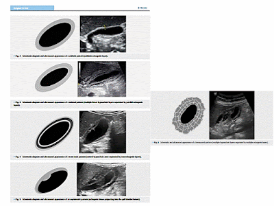
 Dengue fever is one of the most common viral infections with variable clinical presentations and degrees of severity. Gall bladder wall thickening is one of the most common findings in dengue fever. The authors demonstrated four distinct patterns of gall bladder wall thickening in dengue fever patients. The uniform echogenic layer was found to be more common in dengue fever without warning sign, while the honeycomb pattern was more common in severe dengue fever. The research concluded that the patterns of gall bladder wall thickening can be used to diagnose the severity of disease and, when combined with the pattern type in serial ultrasound, may predict the prognosis of the disease.

FURTHER READING:

DÀY VÁCH TÚI MẬT : CHẨN ĐOÁN PHÂN BIỆT

Thứ Hai, 22 tháng 1, 2018

TƯƠNG LAI SIÊU ÂM ĐÀN HỒI GAN trong GAN HỌC


Bài báo đầu tiên về đàn hồi gan với nhiều kết quả khả quan được công bố hơn 10 năm. Bài báo đầu tiên này từ Âu châu và  theo đó của Á châu, đặc biệt dựa vào đàn hồi thoáng qua (TE, transient elastography). So sánh TE với sinh thiết gan, vốn và đang là  tiêu chuẩn vàng đánh giá gan. Trong giai đoạn này việc điều trị viêm gan mạn do hạn chế của thuốc [đặc biệt là pegylated interferon]  với kết quả không được tốt. Quyết định điều trị dựa vào đa số ca được sinh thiết gan. Mới, là gần đây có nhiều thuốc trị viêm gan mạn có hiệu quả  như direct acting agents (interferon-free treatment) cho viêm gan  C mạn  hay  nucleoside/nucleotide analogs (cho viêm gan  B mạn).
Vấn đề khởi phát hiện nay là khi nào cần sinh thiết gan trước khi điều trị. Hầu hết bệnh nhân được điều trị sẽ lành bệnh hay sạch siêu vi mà không cần đặt nặng việc sinh thiết gan. Viễn cảnh đánh giá xơ hóa gan đã thay đổi trong gan học hiện nay: từ sinh thiết gan tất cả đến chỉ sinh thiết giới hạn những ca không rõ ràng. Kỹ thuật siêu âm đàn hồi hay test sinh học [đơn giản hay phức tạp] đã được dùng đánh giá xơ hóa gan trước và sau điều trị. Chiến lược này khởi sự ở Âu châu, nhưng mới đây, theo sau chuẩn y máy siêu âm của FDA, cũng được áp dụng ở Mỹ.
Vấn đề khác là khi nào sinh thiết gan trong thực hành hàng ngày. Câu trả lời của nhiều bác sĩ [và con số đang gia tăng] là  khi không rõ ràng. Không rõ ràng có nghĩa là khi tham khảo các phương pháp không xâm lấn đánh giá độ nặng của xơ hóa gan, chủ yếu là kết quả không tương hợp giữa sinh học và siêu âm đàn hồi. Các trường hợp khác cần phải sinh thiết gan là các trường hợp đặc biệt như viêm gan tự miễn, bệnh lý gan không do rượu, v.v..
Test sinh học giúp thông tin về  độ nặng xơ hóa gan , hoạt động / và hoặc gan thấm mỡ. Bằng cách kết hợp với siêu âm đàn hồi, có thể biết độ nặng của gan thấm mỡ và độ cứng gan như chỉ dấu [marker] của giai đoạn gan hóa xơ. Quyết định ra sao giữa kết quả  sinh học và đàn hồi ? Điều này tùy thuộc vào chuyên gia tại chỗ, khả năng máy siêu âm thực hiện được siêu âm đàn hồi và giá cả của các kỹ thuật tại chỗ.
Hiên thời có nhiều dòng máy siêu âm đàn hồi. Kỹ thuật đàn hồi được phân thành đàn hồi sóng biến dạng (shear wave elastography (SWE) [transient elastography (TE), point SWE và 2-dimensional SWE (2D-SWE)]) và đàn hồi căng [strain elastography]. Các kỹ thuật SWE hiện đang sẵn để sử dụng lâm sàng.
Ngoài ra cộng hưởng từ đàn hồi [magnetic resonance elastography (MRE)] được Mỹ sử dụng mạnh. Do có nhiều kết quả rất tốt, MRE được sử dụng thường ngày [ít nhất tại Mỹ]. Làm sao chọn giữa siêu âm đàn hồi hay MRE ?. Giá cả có thể là yếu tố quyết định. Đối với bác sĩ siêu âm thì siêu âm đàn hồi SWE đủ hiện đại nên thường sử dụng. Với kỹ thuật này việc trả lời kết quả gan thấm mỡ [ít nhất trung bình hoặc nặng] chỉ dưới 5 phút. Radiologist có thể khó quyết định siêu âm hay MRE. Tuy nhiên các nhà bệnh học gan dùng máy siêu âm  và có máy siêu âm SWE thì lựa chọn dễ dàng hơn. Họ sẽ dùng sóng âm.
Lợi ích của chiến lược là gì? Là chẩn đoán tiếp cận [“point of care” diagnosis]. Bệnh nhân đến khám với vấn đề [như là gan tôi có hư hoại nặng không?] và nhận ngay kết quả định thời [staging] bệnh lý gan chính xác đầy đủ  trong ít phút. Đừng quên, nếu phát hiện có khối trong gan, siêu âm có chất tương phản [contrast-enhanced ultrasound (CEUS)] giúp chẩn đoán với xác suất cao.

Do đó, nhìn về tương lai của đàn hồi gan, chẳng bao lâu các nhà bệnh học gan có thể siêu âm đàn hồi gan cho bệnh nhân [đánh giá gan thấm mỡ, cấu trúc gan, bề mặt gan, dấu hiệu cao áp tĩnh mạch cửa, báng bụng, khối u, v.v..], theo dỏi bằng SWE để đánh giá độ cứng gan như là chỉ dấu của độ nặng hoá xơ gan. Nếu phát hiện u gan thì trong cùng lần khám sẽ thực hiện siêu âm với chất tương phản.

-----------------------------------------------------------------------------------------------
More than 10 years ago, the first papers on liver elastography were published, showing its good results. The first publications were European followed by Asian ones, and were focused especially on transient elastography (TE). They compared TE to liver biopsy which was, and still is, considered to be the “gold standard” of liver evaluation. During that time, the treatment of chronic hepatitis was limited to drugs with rather poor results (especially pegylated interferon). Decision for treatment was based in the vast majority of cases on liver biopsy. New, very potent drugs became available in recent years for viral chronic hepatitis: direct acting agents (interferon-free treatment) for chronic hepatitis C or nucleoside/nucleotide analogs (for chronic hepatitis B).
The question that arises now is whether it is necessary to perform a liver biopsy before treatment. Almost all patients that are treated will be healed or will become aviremic, with no regard to liver fibrosis severity.
The perspective regarding the assessment of liver fibrosis severity has changed in modern hepatology: from liver biopsy in all cases, to only a limited number of biopsies in unclear cases. Ultrasound-based elastographic methods or biological tests (simple or complex) are used to assess fibrosis before and after treatment  . This strategy started in Europe, but very recently, with the FDA approval of elastographic systems, it began to be used also in the USA.
Another question that arises is when to perform liver biopsy in daily practice. For many clinicians (and the number is increasing) the answer is: when something is unclear. What does unclear mean when referring to noninvasive techniques to evaluate liver fibrosis severity: Mainly discordant results between biological and elastographic methods. Another situation in which liver biopsy is recommended is special cases of autoimmune hepatitis, non-alcoholic liver diseases, etc.
Biological tests can provide information regarding the severity of fibrosis, activity/and or steatosis. By combining ultrasound evaluation with liver elastography, we can obtain information regarding steatosis severity and regarding liver stiffness as a marker of fibrosis stage. How is a decision between biological tests and elastography made? This probably depends on the local expertise, the availability of ultrasound machines able to perform elastography, and on the local cost of the available methods.
At present, there are many ultrasound systems that can perform liver elastography. Ultrasound-based elastography techniques can be divided into shear wave elastography (SWE) [transient elastography (TE), point SWE and 2-dimensional SWE (2D-SWE)] and strain elastography   . SWE methods are probably currently ready to be used in clinical practice    .
On the other hand, magnetic resonance elastography (MRE) is strongly promoted by radiologists from the USA. As a result of the very good results of this method, it will soon be used in daily practice (at least in the USA). How is a selection between ultrasound-based elastography and MRE made? The cost will probably be the deciding factor. For clinicians using an ultrasound machine that is modern enough to have an SWE module, they will always use ultrasound-based elastography. With this technique, answers regarding steatosis (at least moderate and severe) and liver stiffness can be obtained in less than 5 min. For radiologists it will probably be more difficult to decide between MRE and ultrasound-based elastography. However, for hepatologists using ultrasound and having modern machines (with SWE), the choice is not difficult at all. They will use ultrasound waves.
What is the advantage of this strategy? The advantage is “point of care” diagnosis. The patient comes with a problem (such as “is my liver severely damaged?”) and receives an answer in only a few minutes with sufficiently accurate staging of his/her liver disease. Don’t forget that if a mass is discovered in the liver by ultrasound examination, contrast-enhanced ultrasound (CEUS) will be able to provide a diagnosis with high probability.
Thus, with respect to the future of liver elastography, I believe that very soon every hepatologist will perform clinical ultrasound for his/her patients (assessing steatosis, liver structure, liver surface, signs of portal hypertension, ascites, masses, etc.), followed by SWE for the evaluation of liver stiffness as a marker of fibrosis severity. If a mass is discovered, a CEUS evaluation can be performed in the same session.

Footnotes

Conflict of interest The authors declare that they have no conflict of interest.

References

1. EASL Recommendations on Treatment of Hepatitis C 2015. J Hepatol 2015. 63:199–236. [PubMed]
2. Bamber J, Cosgrove D, Dietrich C F et al. EFSUMB guidelines and recommendations on the clinical use of ultrasound elastography. Part 1: Basic principles and technology. Ultraschall in Med. 2013;34:169–184.[PubMed]
3. Shiina T, Nightingale K R, Palmeri M L et al. WFUMB guidelines and recommendations for clinical use of ultrasound elastography: Part 1: basic principles and terminology. Ultrasound Med Biol. 2015;41:1126–1147. [PubMed]
4. Cosgrove D, Piscaglia F, Bamber J et al. EFSUMB guidelines and recommendations on the clinical use of ultrasound elastography. Part 2: Clinical applications. Ultraschall in Med. 2013;34:238–253. [PubMed]
5. Ferraioli G, Filice C, Castera L et al. WFUMB guidelines and recommendations for clinical use of ultrasound elastography: Part 3: Liver. Ultrasound Med Biol. 2015;41:1161–1179. [PubMed]
6. EASL-ALEH Clinical Practice Guidelines: Non-invasive tests for evaluation of liver disease severity and prognosis. J Hepatol. 2015;63:237–264. [PubMed]

Articles from Ultrasound International Open are provided here courtesy of Thieme Medical Publishers

Thứ Ba, 16 tháng 1, 2018

CÔNG THỨC GOECKE 2: SIÊU ÂM ĐO THỂ TÍCH TRÀN DỊCH MÀNG PHỔI

Ultrasonography is safe, cheap, and convenient route, and is easy to use in most patients, except in very young children. It is also convenient to use in patients who are in the intensive care unit. For daily clinical applications, a simple single measurement of the volume of pleural effusion is preferable to a formula requiring multiple measurements.
While the two erect formulae gave quite accurate measurements, the ultrasonographically 
estimated volume correlated most closely with the actual volume drained when the erect 2 (Goecke 
2) formula was used. It is hoped that these findings will be useful for the accurate quantification of 
pleural effusion in daily clinical practice.