Tổng số lượt xem trang

Thứ Năm, 12 tháng 7, 2018

Dense breast tissue translates into higher cancer odds.


By Kate Madden Yee, AuntMinnie.com staff writer
June 26, 2018 -- A large study out of Norway found that women with dense breast tissue do have higher odds of both screen-detected and interval breast cancer -- and that automated breast density measurement tools can accurately categorize tissue density, according to a study published online June 26 in Radiology.
The findings suggest that automated density measurement tools offer an effective alternative to subjective density assessments, and they point to the need to continue notifying women of their tissue density and improving supplemental screening methods, wrote Dr. Liane Philpotts of Yale School of Medicine in an editorial that accompanied the research.
"This study is important for two main reasons," Philpotts wrote. "First, [it] provides validation that automated means of density classification can correctly identify a percentage of women with dense tissue. Second, women with dense tissue have poorer performance at screening mammography. This study lends support to the density notification movement, along with redoubling efforts toward optimizing methods of supplemental screening."
Density matters
Breast density has been under examination in recent years as researchers have investigated its effects on screening mammography and, in light of mammography's limitations with dense tissue and a grassroots density notification movement, sought to develop alternative imaging options. All along, the issue of how to characterize breast tissue -- whether through radiologists' subjective judgments based on the BI-RADS scale or through automated tools -- has continued to be debated.
"No reference standard exists for breast density determination," wrote the team led by Dr. Nataliia Moshina, PhD, from the Cancer Registry of Norway in Oslo. "[And although for example, in] the United States, more than half the states have enacted breast density notification legislation ... [which] mandates that women should be informed about their breast density or that additional imaging could be beneficial ... supplemental screening for women with dense breasts is not currently recommended by any major societies or organizations."
Moshina and colleagues sought to assess the effectiveness of automated volumetric breast density measurement software in identifying women with dense tissue, as well as to examine how dense breast tissue affects screening performance and outcomes. The group used data from BreastScreen Norway, a screening initiative coordinated by the Cancer Registry of Norway, and included 107,949 women between the ages of 50 and 69 who underwent 2D screening mammography between January 2007 and December 2015, resulting in 307,015 exams. Moshina's team tracked recall, biopsy, and rates of screen-detected and interval cancer, along with sensitivity, specificity, and histopathologic tumor characteristics.
Breast density was assessed with automated software from Volpara Solutions, which uses a four-category scale considered comparable to BI-RADS density categories:
  • Density grade 1: ≤ 4.5% density (BI-RADS 1, fatty tissue)
  • Density grade 2: 4.5% to 7.49% density (BI-RADS 2, scattered fibroglandular tissue)
  • Density grade 3: 7.5% to 15.49% density (BI-RADS 3, heterogeneously dense tissue)
  • Density grade 4: ≥ 15.5% density (BI-RADS 4, extremely dense tissue)
Of the total number of breasts screened, 28% were classified as dense. The researchers found that dense breast tissue translated into higher recall rates, lower mammographic sensitivity, larger tumor size, and more lymph node-positive disease.
Characteristics of screen-detected cancers by density category
MeasureNondense tissue (volumetric density < 7.5%)Dense tissue (volumetric density ≥ 7.5%)p-value
Biopsy rate1.1%1.4%< 0.0001
Positive lymph node18%24%0.02
Rate of interval cancer (per 1,000 exams)1.22.8< 0.0001
Rate of screen-detected cancer (per 1,000 exams)5.56.70.0001
Recall rate2.7%3.6%< 0.0001
Sensitivity81.6%70.8%< 0.0001
Specificity97.9%97%< 0.0001
Tumor diameter (mean)15.1 mm16.6 mm0.009
The adjusted odds of screen-detected breast cancer were 1.37 times higher in women with dense breasts than in those with nondense tissue. In addition, the odds of interval cancer were 2.93 times higher in women with dense tissue than in their nondense counterparts, the researchers found.
"These findings suggest that mammographic density impacts the performance of breast cancer screening and the results show worse outcome for women with dense breasts," corresponding author Solveig Hofvind, PhD, told AuntMinnie.com via email. "The study corroborates previous findings on the association of high breast density and less favorable tumor characteristics, including tumor size and lymph node involvement."
Dealing with density
Using automated volumetric density assessment software helped Moshina's team confirm that women with dense breast tissue have higher recall and biopsy rates and higher odds of screen-detected and interval breast cancer, as well as larger tumors and more positive lymph nodes. Although the higher recall and biopsy rates were likely due to interpretation challenges, they noted that these rates were still substantially lower than rates in the U.S., which are usually around 10%.
The researchers also emphasized that the relatively small difference in biopsy rates between women with dense and nondense breast tissue is "clinically relevant for a screening program that covers about 85% of the 600,000 women in [Norway's] target population." And the finding that mammography is less sensitive in dense tissue underlines the need to find effective supplemental imaging.
"The lower sensitivity of mammography screening among women with dense breasts indicates that mammographic screening is less effective for [them]," the group wrote. "This is because of a higher rate of interval breast cancers with less favorable tumor characteristics compared with screen-detected breast cancers."
The study suggests that automated volumetric density categorization software is a helpful tool for identifying women at higher risk of breast cancer, and it could improve the way women with dense tissue are identified and tracked, Philpotts concluded in her editorial.
"Automated volumetric determination could better standardize density compared with subjective methods and provide greater confidence that this group of women could consistently be identified," she wrote. "This should also help radiology practices to be open to adopting methods to screen such women. Breast density is here to stay, and it is in everyone's best interest to embrace understanding and optimization of breast imaging practice to best address the needs of women with dense tissue."

Thứ Bảy, 30 tháng 6, 2018

NHÂN CA THOÁT VỊ TRƯỢT BÀNG QUANG VÀO BÌU TRÁI

Thoát vị bàng quang vào bìu hiếm gặp. Thường là thoát vị bẹn trượt (sliding inguinal hernia), có nhiều nguyên nhân, nhưng ca 501 Medic chỉ do thành bụng bẹn T yếu, trong khi y văn cho thấy là do biến chứng sau mổ tiền liệt tuyến với vùng bẹn yếu, hay giảm trương lực bàng quang thứ phát do u xơ tiền tuyến hay bế tắc đường tiểu. Triệu chứng điển hình là tiểu 2 lần: lần đầu tiểu ngẩu nhiên bình thường, kế đó ép khối vùng bẹn và đi tiểu tiếp tục.
Chẩn đoán  trước mổ chủ yếu là siêu âm và chụp cản quang bàng quang ngược dòng, với hình bàng quang thoát vị dạng quả tạ [dumbbell shaped urinary bladder];  chẩn đoán đúng gíup tránh làm rách bàng quang khi mổ khâu thoát vị bẹn [herniorhapphy].


J Radiol Case Rep. 2009;3(2):7-9. doi: 10.3941/jrcr.v3i2.91. Epub 2009 Feb 1.

Cystogram with dumbbell shaped urinary bladder in a sliding inguinal hernia.

Abstract

Sliding inguinal hernias present with various symptoms and these are usually direct inguinal hernias containing various abdominal viscera. Case reports and series have been published with various organs and rare organs being part of the hernia. Urinary bladder is a known content of sliding hernias. This case report emphasizes this aspect in a picturesque manner and the importance of radiological investigations for pre-surgical evaluation.

KEYWORDS:

Bladder herniation; Cystogram; Inguinal hernia; Prostatic hypertrophy; Sliding hernia


 [Bladder hernia]. Ann Ital Chir. 1995 May-Jun;66(3):363-9.
 [Article in Italian]
Abstract
A case of bladder hernia in a 61 years old patient affected by benign prostatic hypertrophy is presented. Pre-operative diagnosis was made by cystography. After an adenomiomectomy of the prostate, the patient underwent the resection of the herniated bladder which gave the bladder its normal shape with only a slight reduction of its capacity. Inguino-scrotal bladder hernias are very rare; recognized predisponing factors are weakening of muscular and connective structures of the inguinal canal, and bladder hypotonia secondary to urethro-prostatic obstruction.
These hernias, according to the anatomical position of the hernial sac, bladder and peritoneum, are classified in paraperitoneal (most frequent), intraperitoneal and extraperitoneal. The typical symptom of this disease is the two-stage micturition: the patient after a first spontaneous voiding, presses the mass and voids again. Other than cystography, useful diagnostic means are urography and cystoscopy which may confirm the diagnosis and rule out associated urinary disease.
The treatment consists of either simple reduction of the bladder hernia, if the hernia is small, or resection of the herniated portion of the bladder, if the hernia is large or is associated with other diseases (e.g. tumors). Bladder resection is then followed by closure of the bladder wall in two layers and by inguinal hernia repair.
Actas Urol Esp. 1999 Jan;23(1):79-82.
[A massive hernia of the bladder into the scrotum. A report of a case].
[Article in Spanish]
Abstract
The hernia of the bladder in the scrotum is a highly uncommon observation. From the clinical standpoint the usual manifestation is a two-stroke voiding. The recommended urological examinations to reach a diagnosis are ultrasound, endovenous urography, retrograde urethrocystography and cystoscopy. Management includes the de-obstruction of the lower urinary tract, if present, resection of associated peritoneum, resection or reduction of the vesical hernia and repairment of inguinal path. The case contributed corresponds to a vesical hernia in a 72-year-old patient, with no obstructive cause, that was treated surgically by resection of the herniated bladder, with good morphological and functional results.
 1997 May;21(5):514-8.

[Vesico-scrotal hernia. Report of a clinical case].

[Article in Spanish]

Abstract

Incidental discovery of vesical hernias during herniorrhaphy is quite common. In such cases, patients are usually asymptomatic since the hernia portion is small and easily repairable. On the other hand, vesical solid protrusion to the scrotum is quite unusual, and is generally found associated to obstructive urinary symptoms. Management involves basically the correction of any associated obstructive conditions, correction of the vesical hernia and herniorrhaphy.

US first for Assessing Acute Appendicitis


By AuntMinnie.com staff writers
June 22, 2018 -- Ultrasound, CT, and MRI are all viable supplemental imaging modalities for assessing acute appendicitis after initial ultrasound, according to a study published online June 19 in Radiology.

The team led by Dr. Kevin Eng conducted a literature search on Medline and Embase, identifying studies that used surgery or histopathologic exam information alone or in combination with clinical follow-up or chart review to assess the diagnostic accuracy of supplemental imaging modalities for appendicitis.
Studies included the following:
  • For children -- ultrasound: six studies and 548 patients; CT: nine studies and 1,498 patients; MRI: five studies and 287 patients
  • For adults -- ultrasound: three studies and 169 patients; CT: 11 studies and 1,027 patients; MRI: six studies and 427 patients
All three modalities had comparable, high accuracy for diagnosing the disease, both in children and adults, the researchers found.
Pooled sensitivities and specificities of second-line imaging for diagnosing appendicitis
MeasureUltrasoundCTMRI
Sensitivity: Adults83.1%89.9%97.4%
Sensitivity: Children91.3%96.2%89.9%
Specificity: Adults90.9%93.6%97.1%
Specificity: Children95.2%94.6%93.6%
"All three modalities may be valid as second-line imaging in a clinical imaging pathway for diagnosis and management of appendicitis," they concluded.

Thứ Sáu, 29 tháng 6, 2018

SIÊU ÂM ĐẦU CỔ TRONG CẤP CỨU.

https://blogger.googleusercontent.com/img/proxy/AVvXsEjEduRQAhudhXHqKDYVQM9GkKG8du-cSsx2Podlc_2xyk6TMQY04PyqrKfYl44ucJJdYZVLM_soxA57oSsQHPiXzDQ-vlZcLbU-m8P-IF_5j0shlDIc9yACP3F7g45NpnA3l-DTLwDmB8AZ2XmXwpC7IcLBkwrRkg5oGRBc16IQNOFLSRrz0fXP412d=s0-d-e1-ft

Thứ Năm, 28 tháng 6, 2018

SIÊU ÂM ĐÀN HỒI và GAN HOÁ XƠ Ở TRẺ NẶNG CÂN


Nonalcoholic fatty liver disease is the most common chronic liver disease in overweight adolescents. In most cases, the disease will remain stable for decades without causing significant impairment of liver function. If the disease progresses, persistent inflammation in the liver will cause the formation of excess connective tissue - a stage known as liver fibrosis. Eventually, the disease may damage the liver's entire cellular structure, resulting in what is known as 'liver cirrhosis'.
"Time-harmonic elastography is a new, ultrasound-based technology used to measure liver fibrosis. It works without the need for invasive liver biopsies," explains Dr. Christian Hudert, a pediatric gastroenterologist from Charité's Center for chronically sick children (SPZ). Until now, the grading and staging of liver disease has only been possible through the analysis of tissues taken at biopsy. Elastography is capable of visualizing differences in the elasticity of different tissues. In patients with liver fibrosis, excess connective tissue causes the liver to be stiffer than a normal liver and means that liver stiffness constitutes a measure of disease progression.
The trolley on which the patient is positioned during their examination is a special and defining characteristic of Charité's THE technology. This trolley produces vibrational waves, which are not unlike those produced by a massage chair. Using ultrasound technology, these waves are then measured inside the liver tissue, thus providing information on liver stiffness. In contrast to previous elastography techniques, this technology is capable of taking measurements at greater tissue depths, making it particularly suitable for use in obese patients.

This study used the newly-developed technology to examine 67 adolescents with nonalcoholic fatty liver disease. Liver stiffness measurements were used to determine the degree of fibrosis present. Summing up the study's findings, Dr. Hudert explains: "The THE technology was shown to be capable of accurately distinguishing between patients with no fibrosis or mild fibrosis and patients with moderate or advanced fibrosis." Should the technology prove successful in further studies, it may eventually reduce the need for patients to undergo invasive liver biopsies. The THE method is also particularly suitable for use in the long-term monitoring of patients and may help to verify the success of weight loss treatment options as well as their impact on the outcome of liver disease.

Thứ Tư, 27 tháng 6, 2018

Appendicitis and Clinical Ultrasound


Appendicitis and CUS


Abstract

Introduction: Clinical ultrasound (CUS) is highly specific for the diagnosis of acute appendicitis but is operator-dependent. The goal of this study was to determine if a heterogeneous group of emergency physicians (EP) could diagnose acute appendicitis on CUS in patients with a moderate to high pre-test probability.
Methods: This was a prospective, observational study of a convenience sample of adult and pediatric patients with suspected appendicitis. Sonographers received a structured, 20-minute CUS training on appendicitis prior to patient enrollment. The presence of a dilated (>6 mm diameter), non-compressible, blind-ending tubular structure was considered a positive study. Non-visualization or indeterminate studies were considered negative. We collected pre-test probability of acute appendicitis based on a 10-point visual analog scale (moderate to high was defined as >3), and confidence in CUS interpretation. The primary objective was measured by comparing CUS findings to surgical pathology and one week follow-up.
Results: We enrolled 105 patients; 76 had moderate to high pre-test probability. Of these, 24 were children. The rate of appendicitis was 36.8% in those with moderate to high pre-test probability. CUS were recorded by 33 different EPs. The sensitivity, specificity, and positive and negative likelihood ratios of EP-performed CUS in patients with moderate to high pre-test probability were 42.8% (95% confidence interval [CI] [25-62.5%]), 97.9% (95% CI [87.5–99.8%]), 20.7 (95% CI [2.8–149.9]) and 0.58 (95% CI [0.42–0.8]), respectively. The 16 false negative scans were all interpreted as indeterminate. There was one false positive CUS diagnosis; however, the sonographer reported low confidence of 2/10.
Conclusion: A heterogeneous group of EP sonographers can safely identify acute appendicitis with high specificity in patients with moderate to high pre-test probability. This data adds support for surgical consultation without further imaging beyond CUS in the appropriate clinical setting.

Thứ Hai, 25 tháng 6, 2018

TÀI LIỆU THAM KHẢO CƠ XƯƠNG KHỚP DÀNH CHO LỚP SATQ KHÓA 28

DOWNLOAD LINK

GIÁO TRÌNH CƠ XƯƠNG KHỚP WFUMB 2011

Four Common Pathologies of the Hip Region


MusclePremium_CustomerPage_2017_Header.png

There are three common causes of hip injuries: overuse, sports, and aging. If the hips are used excessively or if the hip joint wears down, the risk for injury increases. And, of course, let's not forget that sudden and/or eccentric movements can also cause injury.

Hip-Pathology-1.png

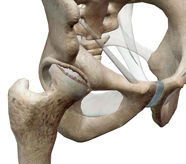
Excessive strain from repetitive pivoting—such as in ice hockey, golfing, or ballet dancing—or direct impact and injury to the hip can tear the tissue of the acetabular labrum. Structural abnormalities and degenerative conditions can also cause a tear or even impingement of the labrum from activities like martial arts, cycling, and horseback riding.

Hip-Pathology-2.png

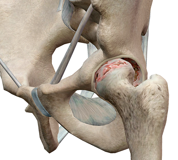
With age and use, the articular cartilage in the hip joint can degenerate. The tissue becomes frayed and rough, exposing the bone surfaces beneath, making movement painful. This condition is diagnosed as osteoarthritis, also known as degenerative arthritis or degenerative joint disease.

Hip-Pathology-3.png

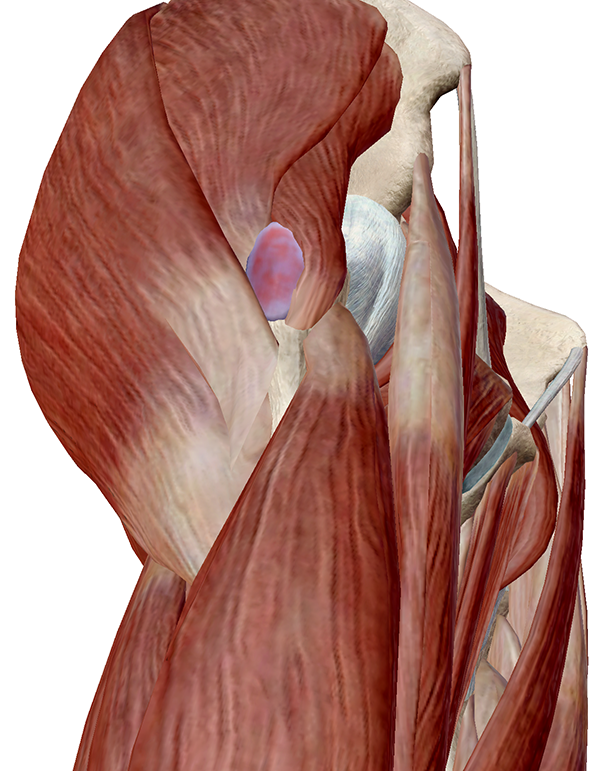
Repetitive stress from activities like extensive running, bicycling, or even standing for long periods of time can irritate and inflame the bursae of the hip, causing sharp pain and tenderness at the point of the hip, usually extending into the thigh.


Hip-Pathology-4.png

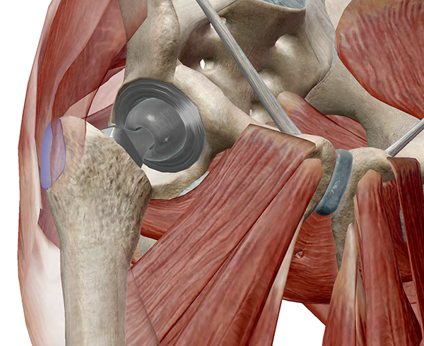
If the pain and stiffness from a damaged hip joint are severe enough or limit mobility, a total hip arthroplasty may be recommended. In hip arthroplasty, the femoral head is removed and replaced with an implant or prosthesis while a metal stem is fitted into the hollow shaft of the femur. A metal ball is then placed on the top of the stem to replace the femoral head. Diseased or damaged articular cartilage is removed from the acetabular surface and replaced with a prosthetic socket. 


ULTRASOUND SCORING US10 of HANDS and WRIST JOINTS in Rheumatoid Arthritis


Thứ Sáu, 22 tháng 6, 2018

I B D in Children and Ultrasound


Abstract

Background

Magnetic resonance enterography (MRE) is the current gold standard for imaging in inflammatory bowel disease, but ultrasound (US) is a potential alternative.

Objective

To determine whether US is as good as MRE for the detecting inflamed bowel, using a combined consensus score as the reference standard.

Materials and methods

We conducted a retrospective cohort study in children and adolescents under 18 years with inflammatory bowel disease (IBD) at a tertiary and quaternary centre. We included children who underwent MRE and US within 4 weeks. We scored MRE using the London score and US using a score adapted from the METRIC (MR Enterography or Ultrasound in Crohn’s Disease) trial. Four gastroenterologists assessed an independent clinical consensus score. A combined consensus score using the imaging and clinical scores was agreed upon and used as the reference standard to compare MRE with US.

Results

We included 53 children. At a whole-patient level, MRE scores were 2% higher than US scores. We used Lin coefficient to assess inter-observer variability. The repeatability of MRE scores was poor (Lin 0.6). Agreement for US scoring was substantial (Lin 0.95). There was a significant positive correlation between MRE and clinical consensus scores (Spearman’s rho = 0.598, P=0.0053) and US and clinical consensus scores (Spearman’s rho = 0.657, P=0.0016).

Conclusion

US detects as much clinically significant bowel disease as MRE. It is possible that MRE overestimates the presence of disease when using a scoring system. This study demonstrates the feasibility of using a clinical consensus reference standard in paediatric IBD imaging studies.

Keywords

Adolescents Children Diagnostic accuracy Inflammatory bowel disease Magnetic resonance imaging Reproducibility Ultrasound 

Thứ Năm, 21 tháng 6, 2018

Elastography of the GI Tract.

Odd Helge Gilja, Prof., MD, PhD National Centre for Ultrasound in Gastroenterology Haukeland University Hospital, Bergen, and Department of Clinical Medicine, University of Bergen, Norway.

Elastography is a relative new technique that depicts the stiffness of tissue and is already used in clinical practice for several indications.

Guidelines and recommendations of the different techniques and clinical applications was published by EFSUMB. Elastography and strain imaging has also been suggested as a tool for assessing diseases of the gastrointestinal tract.
The bowel wall is a thin structure, which does not make it the ideal organ to be studied with elastography. However, pathology of the GI tract such as inflammation or tumour causes bowel wall thickening and often reduces motility and luminal contents in the affected area which may facilitate strain imaging. There is evidence for the use of elastography in endorectal ultrasonography, but the evidence for transabdominal elastography of the bowel is sparse. However, some recent studies suggest that it can be used to differentiate between fibrotic and inflammatory stenosis in Crohn’s disease. In patients with a stricture of the bowel and resultant bowel obstruction, it is important to determine if there is active inflammation at the site of stricture or if the obstructed segment is fibrotic. Using elastography, the active inflammatory components will be softer whereas the fibrotic stricture will appear stiffer. Accordingly, one may apply elastography to evaluate stiffness of the stenotic area, thus providing more clues to the fibrous content of the stricture. In conclusion, elastography may become a relevant clinical tool, but we need more studies to determine its usefulness in the management of patients with GI diseases.

References: 1. Bamber J, Cosgrove D, Dietrich CF, Fromageau J, Bojunga J, Calliada F, Cantisani V, Correas JM, D’Onofrio M, Drakonaki EE, Fink M, Friedrich-Rust M, Gilja OH,Havre RF,JenssenC,KlauserAS,Ohlinger R, Saftoiu A,SchaeferF, SporeaI,PiscagliaF. EFSUMBGuidelinesand Recommendations on the Clinical Use of Ultrasound Elastography. Part 1: Basic Principles and Technology. Ultraschall Med. 2013 Apr;34(2):169-84. http://dx.doi.org/10.1055/s-00331335205. Epub 2013 Apr 4. 2. Cosgrove D, Piscaglia F, Bamber J, Bojunga J, Correas JM, Gilja OH, Klauser AS, Sporea I, Calliada F, Cantisani V, D’Onofrio M, Drakonaki EE, Fink M, Friedrich-Rust M, Fromageau J, Havre RF, Jenssen C, Ohlinger R, Saftoiu A, Schaefer F, Dietrich CF. EFSUMBGuidelines and Recommendations on the Clinical Use of Ultrasound Elastography.

Thứ Tư, 20 tháng 6, 2018

Ultrasound links meat allergen to heart disease


By Kate Madden Yee, AuntMinnie.com staff writer
June 15, 2018 -- Using intravascular ultrasound (IVUS), researchers from the U.S. National Institutes of Health (NIH) have found a link between sensitivity to an allergen in red meat and plaque buildup in the heart's arteries, according to a study published online June 14 in Arteriosclerosis, Thrombosis, and Vascular Biology.
Only recently have scientists identified the main allergen in red meat, called galactose-alpha-1,3-galactose, or alpha-Gal, wrote a team led by Dr. Coleen McNamara from the University of Virginia Health System. It's also been discovered that the lone star tick, found predominately in the southeastern U.S., sensitizes people to this allergen when it bites them, boosting the incidence of meat allergies in this area.
Researchers have suspected for some time that allergens can trigger immunological changes that may be associated with plaque buildup and artery blockages. In their study, McNamara and colleagues showed that a type of antibody (immunoglobulin) specific to the alpha-Gal allergen was associated with higher levels of arterial plaque.
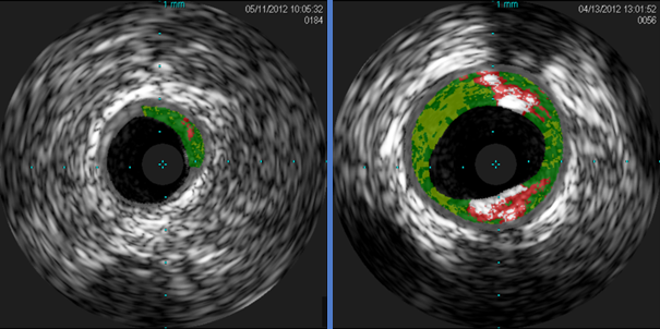
The investigators analyzed blood samples from 118 adults and found antibodies to alpha-Gal in 26%. Next, they used intravascular ultrasound (IVUS) to analyze the quantity of plaque in the blood samples. The amount was 30% higher in the alpha-Gal-sensitized patients than in the nonsensitized patients.
Cross-sectional IVUS images of coronary arteries
Cross-sectional IVUS images of coronary arteries. Plaque buildup (colored areas) in an artery from a patient who lacks sensitivity to the red meat allergen (left) is much lower than plaque levels in an artery from a patient with sensitivity to the allergen (right). Images courtesy of Dr. Angela Taylor of the University of Virginia Health System.
"This novel finding from a small group of subjects from Virginia raises the intriguing possibility that allergy to red meat may be an underrecognized factor in heart disease," McNamara said in a statement released by the NIH. "These preliminary findings underscore the need for further clinical studies in larger populations from diverse geographic regions and additional laboratory work."