Tổng số lượt xem trang

Chủ Nhật, 26 tháng 8, 2018

Vascular Ultrasound and Multiparametric Ultrasound


Siêu âm có truyền thống được coi là phương thức tạo hình đầu tiên để sàng lọc, đánh giá chẩn đoán và điều trị theo dõi và tiến triển bệnh lý mạch máu, bao gồm cả động mạch và tĩnh mạch cả hệ thống. Dù có bản chất dung nạp tốt, tính sẵn sàng rộng và chi phí thấp, trên tất cả, siêu âm có lợi thế là cung cấp cho bác sĩ những thông tin lâm sàng quan trọng liên quan đến bệnh nội mạch và bệnh ngoài mạch. Siêu âm có tiềm năng không chỉ mô tả giải phẫu các mô mà còn để tiếp cận chức năng sinh lý  bằng cách đánh giá đặc điểm lưu lượng máu trong thời gian thực..

Vai trò cơ bản của siêu âm thậm chí còn mở rộng với việc giới thiệu các kỹ thuật mới như siêu âm với chất tăng cường tương phản, siêu âm đàn hồi mô và siêu âm 3D và kết hợp vào thực hành lâm sàng. Mục đích của tổng quan này là trình bày và thảo luận một số tiến bộ mới nhất trong lĩnh vực siêu âm mạch máu nhằm minh họa cách thức áp dụng siêu âm đa năng [multiparametric] vào thực hành lâm sàng hàng ngày nhằm đáp ứng nhu cầu của bệnh nhân. Bệnh lý và các ứng dụng được thảo luận bao gồm bệnh động mạch cảnh và động mạch chủ, tĩnh mạch cửa và bất thường tĩnh mạch ngoại vi.


Máy siêu âm với công nghệ hiện đại tích hợp  đã làm cho việc áp dụng siêu âm với chất 
tăng cường tương phản, đàn hồi mô  và siêu âm 3D với các tùy chọn khả thi, sẵn sàng 
đóng góp vào hiệu suất chẩn đoán của kỹ thuật siêu âm.

Thứ Bảy, 25 tháng 8, 2018

Ultrasound Could Improve Vascular Disease Detection.


By AuntMinnie.com staff writers

August 21, 2018 -- Ultrasound could improve the early detection of vascular disease, perhaps even before patients develop symptoms, according to a preclinical study published August 7 in the journal Ultrasound.
Researchers from the University of Leicester in the U.K. used ultrasound and semiautomatic image processing in mice to investigate the progression of abdominal aortic aneurysm and atherosclerosis. Ultrasound is used for a wide range of diseases and offers real-time information on the ability of arteries to expand and contract.
First author Dr. Justyna Janus and colleagues found changes in the ability of the mice's blood vessels to pulsate as the disease state worsened, suggesting that ultrasound could help in the early detection of reduced vascular function.
"Our study ... suggests that the ultrasound methods we employed can detect early changes in blood vessel function that can serve as a marker for detection of disease," co-author Dr. Mike Kelly said in a statement released by the university.

Thứ Tư, 22 tháng 8, 2018

Ultrasound Imaging Gauges Muscle Tightness after Stroke


August 22, 2018 -- Ultrasound strain imaging can be an effective tool for assessing poststroke muscle spasticity or tightness, a condition that affect two-thirds of stroke survivors, according to a study published in the August issue of the Journal of Ultrasound in Medicine.
The effects of spasticity on muscle after a stroke can include contractures, limited range of joint motion, and pain. Spasticity manifests most often around the elbow, the wrist, and the ankle, but it can also affect other parts of the body, including the biceps brachii muscle, or elbow flexors.
There hasn't been an effective way to measure spasticity to determine which interventions are best for patients after a stroke, wrote a team led by Dr. Jing Gao of Weill Cornell Medicine in New York City.
"To date, this process [of measuring spasticity] remains challenging because of the lack of a reference standard. ... It would be ideal to have a noninvasive imaging technique to quantify the mechanical properties and dynamic movement of spastic muscle to assist clinicians in the diagnosis of spasticity, monitoring of disease progression, and evaluation of treatment response in stroke rehabilitation," the group wrote.
Ultrasound strain imaging has been shown to be useful for assessing the stiffness of skeletal muscle, but little is known about using the technique to assess skeletal muscle dynamic motion. Gao and colleagues sought to evaluate the feasibility of strain imaging for assessing spasticity in the biceps of stroke survivors (J Ultrasound Med, August 2018, Vol. 37:8, pp. 2043-2052).
The study included eight healthy volunteers and seven stroke survivors recruited between February and July 2017; the investigators gathered ultrasound strain imaging data from all participants.
They found a significant increase in muscle stiffness, represented by a decrease in muscle axial strain in the spastic biceps brachii muscles compared with the nonspastic muscles. The researchers also found decreased lengthening and shortening capability (i.e., longitudinal strain ratio) and tissue velocity in the spastic muscles versus the nonspastic ones.
Ultrasound strain imaging measures for biceps brachii muscle
MeasureNonspastic bicepsSpastic biceps
Axial strain ratio (90°)4.873.13
Axial strain ratio (0°)4.022.65
Longitudinal strain ratio (90° to 0°)5.793.12
Longitudinal strain ratio (0° to 90°)6.563.25
Tissue velocity (90° to 0°)2.161.33
Tissue velocity (0° to 90°)-2.09-0.95
The study suggests that ultrasound strain imaging could help clinicians better assess a patient's poststroke spasticity and perhaps better manage the condition, Gao and colleagues concluded.
"Ultrasound strain imaging is a useful imaging tool for determining increased stiffness and decreased dynamic displacement in spastic biceps brachii muscles by assessing the axial strain, longitudinal strain, and tissue velocity of the muscle," they wrote. "These noninvasive strain imaging markers may have potential in improving the point-of-care management of chronic poststroke spasticity."

Thứ Ba, 21 tháng 8, 2018

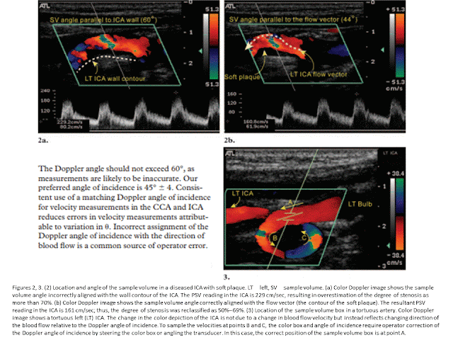
SIÊU ÂM CÁC ĐỘNG MẠCH CẢNH


https://pubs.rsna.org/doi/10.1148/rg.25604501


Conclusions

Carotid US offers a noninvasive evaluation of the extracranial neck portions of the carotid and vertebral arteries for atherosclerotic disease. Standardized technical parameters, scanning methods, Doppler analysis, and interpretation enhance the accuracy and reproducibility of the results.


Abstract

Ultrasonography (US) of the carotid arteries is a common imaging study performed for diagnosis of carotid artery disease. In the United States, carotid US may be the only diagnostic imaging modality performed before carotid endarterectomy. Therefore, the information obtained with carotid US must be reliable and reproducible. Technical parameters that can affect the accuracy of carotid US results include the Doppler angle, sample volume box, color Doppler sampling window, color velocity scale, and color gain. Important factors in diagnosis of atherosclerotic disease of the extracranial carotid arteries are the intima-media thickness, plaque morphology, criteria for grading stenosis, limiting factors such as the presence of dissection or cardiac abnormalities, distinction between near occlusion and total occlusion, and the presence of a subclavian steal. Challenges to the consistency of carotid US results may include lack of a standard protocol, poor Doppler technique, inexperience in interpretation of hemodynamic changes reflected in the Doppler waveform, artifacts, and physical challenges. Hindrances in the classification of problematic carotid artery stenoses may be overcome by following a standard protocol and optimizing scanning techniques and Doppler settings.
© RSNA, 2005

Deep Vein Thrombosis [D V T]

Deep vein thrombosis


https://radiopaedia.org/articles/deep-vein-thrombosis

The term deep vein thrombosis (DVT) is practically a synonym for those that occur in the lower limbs. However, it can also be used for those that occur in the upper limbs and neck veins. Other types of venous thrombosis, such as intra-abdominal and intracranial, are discussed in separate articles.


The majority of deep venous thromboses occur in the lower extremities and begin in the soleal veins of the calf. Doppler compression sonography is the imaging modality of choice.

Features include:
  • non-compressible venous segment
  • increased venous diameter: acute thrombus
  • decreased venous diameter: chronic thrombus
  • loss of phasic flow on Valsalva manoeuvre
  • absent colour flow: if completely occlusive
  • increased flow in superficial veins
  • lack of flow augmentation with calf squeeze
  • anechoic thrombus: acute thrombus
  • echogenic thrombus: chronic thrombus

  Key Points

© Springer International Publishing AG 2018

A. Creditt et al., Clinical Ultrasound,  
https://doi.org/10.1007/978-3-319-68634-9_14


  Using point-of-care ultrasound to evaluate patients  in the emergency department has been shown to decrease length of stay when compared with patients who receive studies performed by the radiology department [4].
  Classically the femoral vein lies medial to the femoral artery; however, a common anatomic variant is for the femoral artery to travel anterior to the femoral vein [1].
  Veins collapse easily with pressure. If you cannot visualize a vein, apply less pressure with the transducer.
  Apply direct and even pressure over the vein to assess its compressibility:
  – A false-positive DVT can be diagnosed if uneven pressure is applied, therefore, resulting in an inability to completely compress the vein.
  – This can also occur with patients who have larger body habitus as it is harder to apply direct and even pressure.
  Noncompressible veins can also be seen with chronic DVTs:
  – When a DVT re-cannulates, it can cause the walls of the vein to become stiffer and less responsive to compression.
  Do not rely on visualization of the clot for diagnosis of a DVT as the appearance changes with maturation:
  – If a vein does not completely compress, this is diagnostic of a DVT even if no thrombus is visualized within the lumen [2].
  While a chronic DVT may appear more echogenic with thickened irregular walls, these findings may not be present and it may not be possible to distinguish an acute from a chronic thrombus [3].
  Point-of-care ultrasound for DVT without the use of color Doppler can be less reliable in patients with recurrent DVT:
  – Approximately 50% of ultrasounds can be abnormal 1 year after initial diagnosis of a DVT [5].

Doppler Ultrasonography of the Lower Extremity Arteries

https://www.e-ultrasonography.org/upload/usg-16054.pdf

Doppler ultrasonography of the lower extremity arteries is a valuable technique, although it is less frequently indicated for peripheral arterial disease than for deep vein thrombosis or varicose veins. Ultrasonography can diagnose stenosis through the direct visualization of plaques and through the analysis of the Doppler waveforms in stenotic and poststenotic arteries. To perform Doppler ultrasonography of the lower extremity arteries, the operator should be familiar with the arterial anatomy of the lower extremities, basic scanning techniques, and the parameters used in color and pulsed-wave Doppler ultrasonography.

Doppler US is the only noninvasive technique that does not require contrast enhancement, preparation of the patient before the study, or radiation exposure [1,2]. Doppler US is a good method for screening and follow-up, as well as for the definitive diagnosis of peripheral arterial disease [3-7]. Color Doppler US can easily identify arteries by finding round objects with regular pulsation and can be used to detect stenotic or occluded segments [4,8]. Pulsed-wave Doppler US can show the exact flow velocity of each arterial segment and determine the degree of severity of the stenosis based on an analysis of the pulsed-wave Doppler spectral waveform [9]. Knowledge of the ultrasonographic anatomy of the lower extremity arteries and the corresponding anatomical landmarks is essential for performing Doppler US. In this article, we review the basic scanning techniques of color and pulsed-wave Doppler US for the lower extremity arteries and the spectral analysis of normal and stenotic arteries on pulsed-wave Doppler US.


Conclusion 

Doppler US has been found to distinguish between stenosis with a diameter reduction greater than or less than 50% (corresponding to an area reduction of 70%) with a sensitivity of 77%-82% and a specificity of 92%-98% [18,20-24]. Complete scanning of both lower extremities may require up to 2 hours depending on the operator’s experience [18]. However, if the operator is familiar with the US anatomy of the lower extremity arteries and understands the parameters and Doppler waveforms of Doppler US, accurate diagnostic results can be produced with less scanning time.

Thứ Hai, 20 tháng 8, 2018

SIÊU ÂM ĐỘNG MẠCH THẬN: BÌNH THƯỜNG và HẸP ĐỘNG MẠCH THẬN

ULTRASOUND OF THE RENAL ARTERIES - NORMAL

https://www.ultrasoundpaedia.com/normal-renal-arteries/


There are 2 techniques, direct method and indirect method, used to investigate renal arterial supply. Both ideally are used in conjuction with each other, however, in circumstances where the renal artery is not seen in its entirety then the indirect approach alone can give an indication of vascular disease.


DIRECT ASSESSMENT


ANTERIOR APPROACH

The renal arteries are clearly imaged in B Mode from an anterior,subcostal approach however as it is perpendicular to the ultrasound beam it is not suitable for Doppler assessment. Supernumerary (duplicate) arteries can be seen looking posterior to the IVC in B Mode and Colour in a sagittal plane.


Anterior Approach.
The patient is lying supine and the probe is moved inferiorly and superiorly to identify the
 renal arteries and any supernumerary arteries. Look in B-Mode and Colour Doppler to help
 identify. A spectral analysis is made of the aorta at the level of the renal arteries. The
velocity is taken with an angle for accurate measurement.If an accurate angle cannot be
obtained then another measurement is taken with no angle so it can be compared to the
renal artery at a stenosis site to do a ratio.The aorta is examined for any abdominal aortic
aneurysm.


OBLIQUE APPROACH

By moving the probe to the left of midline and angling toward the patient's right, an acceptable Doppler angle of 60 degrees is achieved. To avoid aliasing set the colour scale high enough so it is minimized. If the scale is too low then it is difficult to determine which vessel is the vein and which vessel is the artery.

Oblique Approach


Oblique Approach.Angling 45 degrees to right renal artery or rolling the patient in a semi left 

decubitus position to avoid the bowel gas and improve the Doppler angle.


Anterior Approach.

The renal arteries are clearly imaged in B Mode from an anterior approach however as it is
 perpendicular to the ultrasound beam it is not suitable for Doppler assessment.

In most individuals, the renal arteries arise from the abdominal aorta immediately distal to
the origin of the superior mesenteric artery (SMA). The right renal artery passes underneath
the inferior vena cava (IVC)  and posterior to the right renal vein.

ANTERIOR v's OBLIQUE


Oblique Approach.Angling 45 degrees to right renal artery.In most individuals, the renal
arteries arise from the abdominal aorta immediately distal to the origin of the SMA. By moving the probe to the right of midline and angling toward the patient's left, an acceptable Doppler angle of
45-60 degrees is achieved.

Normal waveform
A normal waveform obtained from the main renal artery demonstrates a rapid upstroke in
systole and a low resistance waveform with continuous forward flow throughout the cardiac
 cycle. The normal peak systolic velocity of the main renal artery is less than 150 cm/sec.
The resistive index is less than 0.70 .Doppler sampling throughout the entire artery from
the origin to the hilum.

Coronal Approach 
 
Roll the patient into a decubitus position to avoid bowel gas and improve visibility of the renal artery,especially the mid to distal portion.


INDIRECT METHOD

An indirect assessment requires a good colour image to determine the position of the interlobar and interlobular arteries, which in turn will determine the best angle to get an accurate measurement of the RI and AT.


RIs and ATs (RESISTIVE INDEX AND ACCELERATION TIME)


Resistive Ind
ex
The Resistive Index (RI) is easily performed by placing a caliper on the peak systolic velocity and the other caliper on the lowest diastole.The RI is a ratio of peak systolic and end diastolic velocity.

Acceleration Time
The Acceleration Time (AT) is done by placing a caliper on the level at which the gradient begins to rise and finished at the first peak ie the Early systolic Peak (ESP).This should be less than 70ms


Renal Artery Stenosis

Maria R. Bokhari; Syed Rizwan A. Bokhari.


Renal artery stenosis is narrowing of the one or both of renal arteries. It is the major cause of hypertension and according to some reports is the cause of hypertension in 1% to 10% of the 50 million people in the United States. Atherosclerosis or fibromuscular dysplasia most often cause it. Other associated complications of renal artery stenosis are chronic kidney disease and end-stage renal disease.

Duplex ultrasonographic scanning is a noninvasive, relatively inexpensive technique that can be used in patients with any level of renal function. However,  it is operator dependent and has variable sensitivity. Two approaches are used to detect RAS with Doppler Ultrasound.
  • Direct Visualization of the Renal Arteries: The first approach involves direct scanning of the main renal arteries with color or power Doppler US followed by spectral analysis of renal artery flow using an anterior or anterolateral approach. Owing to various factors essentially as gas interposition and the anatomy of the left renal artery, a complete examination of both renal arteries can be achieved in only 50%–90% of cases. Signal enhancement can be achieved by administering contrast agents that facilitate visualization of the renal arteries. Four criteria are currently used to diagnose significant proximal stenosis:
1.     An increase in peak systolic velocity in the renal artery (the post-stenotic threshold for significant RAS is 100 cm/sec to 200 cm/sec is reported)
2.     A renal-to-aortic peak systolic velocity ratio of greater than 3.5
3.     Turbulent post-stenotic site
4.     Visualization of the renal artery without any detectable Doppler signal, an observation that signals occlusion.
  • Analysis of Intra-renal Doppler Waveforms: The different segments of kidneys are scanned via trans lumbar approach systematically to detect a stenosis of a segmental or accessory renal artery. Quantitative criteria  proposed for detection of significant RAS:
1.     Acceleration of less than 370 cm/sec to 470 cm/sec. The early systolic acceleration seems to be the best predictor of renal artery narrowing
2.     Acceleration time greater than 0.05 sec to 0.08 sec
3.     Change in the resistive index  (RI) of greater than 5% between the right and left kidneys.  This value should be measured along the initial portion of the systolic rise and avoid including the late compliance peak
4.     A dampened presentation (pulsus tardus) of an intrarenal Doppler waveform indicates stenosis
5.     The presence of an early systolic peak can be interpreted as a sign of normality; however, the absence of an early systolic peak is not necessarily indicative of stenosis.


Chronic kidney disease


Dr Henry Knipe and Dr Bruno Di Muzio et al.

Ultrasound with Doppler examination of intrarenal vessels is usually performed in patients with CKD, and it is common to have a normal exam.
Typical B-mode findings of a long-standing severe CKD (especially stage 5) are 2:
  • reduced renal cortical thickness under 6mm more reliable than length 7

  • reduced renal length
  • increased renal cortical echogenicity 
  • poor visibility of the renal pyramids and the renal sinus
  • marginal irregularities
  • papillary calcifications
  • cysts (see also: acquired cystic kidney disease)

  • Abnormal Doppler findings in these patients are 2:
    • reduced renal vascularity
    • increased resistance index (RI) values (segmental and interlobular arteries)

    Thứ Bảy, 18 tháng 8, 2018

    ONSD and Increased Intracranial Pressure

    Siêu âm qua nhãn cầu để đo đừơng kính bao dây thần kinh thị giác, cách củng mạc 3 mm, là phương pháp phát hiện tăng áp nội sọ hiệu quả nhanh, chính xác, dễ thực hiện và không xâm lấn. Bài tổng quan này kết luận tiềm năng của siêu âm đo bao dây thần kinh thị giác (ONSD) sau nhãn cầu trong lâm sàng tăng áp nội sọ.

    ĐO ĐK DÂY TK THỊ GIÁC SAU NHÃN CẦU TIÊN ĐOÁN TĂNG ÁP NỘI SỌ

    TĂNG ÁP NỘI SỌ: SIÊU ÂM ĐO ONSD


    Thứ Bảy, 4 tháng 8, 2018

    SIÊU ÂM XÁC NHẬN RƯỢU GÂY BỆNH GAN

    Siêu âm bụng  301 ca, tuổi trung bình 46, uống rượu ít nhất 10 năm với lượng 180gr mỗi ngày, ngoài tăng men gan ALT và bilirubin, Fuster và cs  phát hiện gan thấm mỡ, gan to, gan không đồng dạng và  cao áp tĩnh mạch Cửa.
    Gan thấm mỡ không viêm có thể trở lại bình thường hay nhẹ đi nếu ngưng uống rượu.

    Siêu âm  phát hiện bệnh lý gan do rượu trong giai đoạn sớm giúp quyết định ngừng uống rượu. 


    By Kate Madden Yee, AuntMinnie.com staff 
    August 1, 2018 -- Using ultrasound to identify liver damage in alcoholic patients helps inform treatment decisions in this population and may even lead to improved prognoses, according to a study published online in Drug and Alcohol Dependence.

    Liver disease due to alcohol use can manifest in a variety of conditions: steatosis, steatohepatitis, acute alcoholic hepatitis, cirrhosis of the liver, and hepatocarcinoma. Some of these can be reversed or mitigated if the patient quits drinking. For example, steatosis without inflammation can be reversed if drinking is stopped. Once steatohepatitis has been established, liver damage can't be fully reversed, but quitting alcohol can lessen portal hypertension.
    And since alcoholic patients tend to be more vulnerable to hepatitis C, a timely diagnosis can lead to better prognosis and treatment planning, wrote a team led by Dr. Daniel Fuster of Autonomous University of Barcelona in Spain (Drug Alcohol Depend, July 19, 2018).


    Although ultrasound is regularly used to diagnose nonalcoholic steatohepatitis, it's less frequently used for the screening and early detection of liver disease in patients with alcohol use disorder (AUD), the authors noted.
    "New strategies aimed at the early detection and treatment of liver-related conditions are needed for this population. Liver ultrasound is an easy-to-perform technique that is rarely used in asymptomatic patients with AUD," the team wrote. "We aimed to describe ultrasound findings of liver disease in patients seeking treatment for AUD."
    Common cause
    Liver disease is a leading cause of death around the world and can include chronic viral hepatitis, alcoholic liver disease, and nonalcoholic liver disease. But disease related to the consumption of alcohol is on the rise, according to Fuster and colleagues.
    "Both in the U.S. and some European countries, there has been an increase in liver-related mortality due to end-stage liver disease, mostly because of the impact of alcohol consumption," the group wrote.
    For the study, Fuster's team used abdominal ultrasound to identify steatosis (fatty liver), hepatomegaly (liver enlargement), heterogeneous liver, and portal hypertension in 301 patients with median age of 46 and alcohol consumption of 180 grams per day for at least 10 years. The group defined analytical liver injury as at least two of the following:
    • Aspartate aminotransferase (AST) levels between 74 and 300 units per liter (U/L)
    • AST/alanine aminotransferase ratio of greater than 2
    • Total bilirubin of more than 1.2 milligrams per deciliter (mg/dL)
    The group defined advanced liver fibrosis as a Fibrosis-4 (FIB-4) score of 3.25 or higher (scores range from higher   1.45 to higher  3.25).
    Among the patient cohort, prevalence of the hepatitis C virus was 21.2%, AST levels were 42 U/L, and ALT levels were 35 U/L (indicating liver enlargement or mild to moderate fatty liver disease); 16% of patients had analytical liver injury and 24% had advanced liver fibrosis.
    Of all the patients, 77% had at least one ultrasound abnormality, and 45% had at least or more than two. Abnormal ultrasound findings included the following:
    • 57.2% steatosis
    • 49.5% liver enlargement
    • 17% heterogeneous liver
    • 16% portal hypertension
    Patients with hepatitis C had a higher incidence of heterogenous liver and portal hypertension compared with those without the infection (25.4% versus 14.5% and 27.1% versus 13.8%), the group found.
    "Liver damage is a major driver of disease burden in patients with unhealthy alcohol use, and liver disease is often diagnosed in advanced stages," Fuster and colleagues wrote. "Results ... indicate that there is a need for the assessment of liver damage in patients seeking treatment for alcohol use disorder."


    Another tool in the arsenal
    The consumption of alcohol is associated with liver disease progression and increased risk of alcoholic hepatitis, according to Fuster and colleagues. So using abdominal ultrasound in patients seeking treatment for alcohol dependency could be helpful for making clinical decisions and taking appropriate therapeutic action. Perhaps it could even give patients more incentive for quitting drinking, the group wrote.
    "Liver ultrasound appears to capture a significant proportion of mild to moderate abnormalities in patients who do not meet the [analytical liver injury] or [advanced liver fibrosis] criteria," the team concluded. "In this regard, the use of ultrasound to detect early stages of liver disease may promote alcohol cessation."