Tổng số lượt xem trang
Chủ Nhật, 8 tháng 5, 2016
Thứ Sáu, 1 tháng 4, 2016
Thứ Hai, 28 tháng 3, 2016
WHAT MEANS POINT-SHEAR WAVE ELASTOGRAPHY [p-SWE]?
Ultrasound based-elastographic techniques are classified in: strain techniques and shear
wave elastography techniques. Three types of elastographic techniques are included
in the last category: Transient Elastography, point Shear Wave Elastography (pSWE)
and shear wave elastography (SWE) imaging (including 2D-SWE and 3D-SWE).
In the pSWE category two techniques are included: Acoustic Radiation Force Impulse (ARFI) elastography and ElastPQ.
Elastographic Techniques Based on Shear Waves Generated by the Acoustic Beam
These techniques have the advantage of being integrated into
ultrasound systems; thus, conventional sonography, which is advised every 6 to
12 months in patients with chronic liver disease, could also be performed. As
of today, for the assessment of liver stiffness, these techniques are
commercially available in high-end ultrasound systems made by Philips
Healthcare (Bothell, WA; ElastPQ), Siemens Medical Solutions (Mountain View, CA;
Virtual Touch Tissue Quantification [VTTQ]), and SuperSonic Imagine, SA
(Aix-en-Provence, France; ShearWave Elastography [SWE]). These techniques
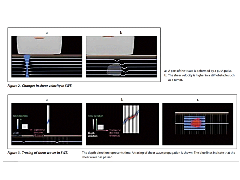
generate shear waves inside the liver by using radiation force from a focused
ultrasound beam. The shear waves are generated near the region of interest in
the liver parenchyma and not on the surface of the body, as happens with
external vibration devices. The ultrasound system monitors shear wave
propagation using a Doppler-like ultrasound technique and measures its
velocity. The shear wave velocity is displayed in meters per second or
kilopascals through the Young modulus. Unlike transient elastography, the
measurements are not limited by the presence of ascites because the ultrasound
beam, which generates the shear waves, propagates through fluids. With the VTTQ
and ElastPQ techniques, the readings of the shear wave speed are made by using
a small sample box (usually 0.5 × 1 cm); thus, a quantitative estimate of liver
stiffness at a single location is obtained (Figures 2 and 3). They have been
categorized as point–shear wave
elastography.The SWE technique is based on an ultrafast ultrasound
imaging approach that allows detailed monitoring of the shear waves in a large
area of liver parenchyma with real-time color-coded elasticity imaging inside a
sample box, and the measurement is obtained by placing a region of interest
inside the sample box (Figure 4). This technique is 2-dimensional
elastography.27 In all of the studies that have assessed the accuracy of the
different devices in staging liver fibrosis, right intercostal access has been
used. The patient is examined in the dorsal decubitus position with the right
arm elevated above the head for optimal intercostal access in a resting respiratory
position. Measurements are performed at least 1.5 to 2.0 cm beneath the Glisson
capsule to avoid reverberation artifacts. In case of physical conditions
affecting the signal to-noise ratio, the Philips and Siemens devices do not
give any measurement. With the SuperSonic Imagine device, a measurement fails
when no/little signals are obtained in the sample box for all of the
acquisitions.
Siemens Technique
(VTTQ)
The first one available was the Siemens technique, which is
commonly referred to as acoustic radiation force impulse in the literature,
which is technically the same force that generates shear waves for all 3
available techniques. Moreover, the term acoustic radiation force impulse is
rather generic and does not identify shear wave–based methods. In fact,
acoustic radiation force impulse push pulses are also used in strain imaging of
other organs, such as the breast and thyroid. In recent years, the diagnostic
accuracy of the VTTQ technology for quantification of liver stiffness, mainly
in patients with chronic hepatitis C, has been investigated in several studies
and a meta-analysis. The technology has shown high interobserver
agreement, with an intraclass correlation coefficient of 0.86. Operator
training does not seem to be required.The cutoff values obtained in a large
meta-analysis were 1.34, 1.55, and 1.80 m/s for significant fibrosis (METAVIR fibrosis
score of F2 or greater), severe fibrosis (METAVIR fibrosis score of F3 or
greater), and cirrhosis (METAVIR fibrosis score of F4), respectively. In this
meta-analysis, which included patients with several etiologies of chronic liver
disease, the diagnostic accuracy was comparable with that of transient
elastography for the assessment of severe fibrosis, whereas higher performance
of transient elastography was seen for significant fibrosis and liver
cirrhosis. In a study by Rizzo et al, the technique was significantly more
accurate than transient elastography for diagnosing significant and severe
fibrosis, whereas this difference was only marginal for cirrhosis.
SuperSonic Imagine
Technique (SWE)
The reproducibility of the SWE method is very high, with
intraobserver intraclass correlation coefficients of 0.95 and 0.93 for an
expert and a novice operator, respectively, and interobserver agreement of
0.88. As for conventional sonography, it is user dependent; thus, it is
recommended that at least 50 supervised scans and measurements should be
performed by a novice operator to obtain consistent measurements. Values
obtained in a small series of healthy participants ranged from 4.92 kPa (1.28
m/s) to 5.39 kPa (1.34 m/s). In a pilot study conducted on 121 patients with
chronic hepatitis C undergoing liver biopsy, the optimal cutoff values were 7.1
kPa (1.54 m/s) for significant fibrosis (METAVIR fibrosis score of F2 or
greater), 8.7 kPa (1.70 m/s) for advanced fibrosis (METAVIR fibrosis score of
F3 or greater), and 10.4 kPa (1.86 m/s) for cirrhosis (METAVIR fibrosis score
of F4), and the technique was more accurate than transient elastography in
assessing significant fibrosis. In another study, with respect to transient
elastography, the technique showed higher accuracy in assessing mild and
intermediate stages of fibrosis.
Philips Technique
(ElastPQ)
The ElastPQ technique was the most recent to enter the
market; thus, only a few studies have been published so far. With this
technique, liver stiffness values in healthy volunteers have been reported to
be less than 4.0 kPa (1.15 m/s). Ling et al found that men had higher
values than women (3.8 ± 0.7 versus 3.5 ± 0.4 kPa, or 1.13 ± 0.48 versus 1.08 ±
0.37 m/s) and liver stiffness was comparable with different probe positions,
examiners, and age groups. In a series that comprised 88 patients with chronic
viral hepatitis and 33 healthy volunteers, the technique compared favorably
with transient elastography in staging liver fibrosis, and healthy volunteers
showed significantly lower values than patients with nonsignificant fibrosis.
Bamber J, Cosgrove D, Dietrich CF, et al. : EFSUMB guidelines and recommendations
on the clinical use of ultrasound elastography, part 1: basic
principles and technology. Ultraschall Med 2013; 34:169–184.
Ferraioli et al: Shear Wave Elastography for Evaluation of Liver Fibrosis, J Ultrasound Med 2014; 33:197–203 199
Thứ Bảy, 26 tháng 3, 2016
Thứ Hai, 14 tháng 3, 2016
Thứ Bảy, 5 tháng 3, 2016
S W ELASTO into PLAQUE IMAGING from E C R 2016
ELASTOGRAPHY OFFERS NEW INSIGHTS INTO PLAQUE IMAGING
ELASTOGRAPHY
OFFERS NEW INSIGHTS INTO PLAQUE IMAGING
Elastography has been used for many years to differentiate
malignant from benign lesions, especially in the breast or liver. Experience in
carotid artery disease is limited, but recent studies have shown that
elastography may help to stratify plaque and reduce the risk of unnecessary
surgery, as a Greek expert will show during a New Horizons session today at the
ECR.
Stroke is one of the leading causes of death in developed
countries; one third of cases are fatal and survival can come with considerable
disabilities. In Europe alone, experts estimate that there are one million new
ischaemic strokes per year and they expect this number to rise by 12% by 2020,
as the population ages1. A wide spectrum of carotid artery diseases can lead to
stroke, but atherosclerosis accounts for a significant percentage – about 20 to
30% of cases. Stenosis is typically a cause for atherosclerosis and is now
being measured using ultrasound in symptomatic patients, who are usually
treated with atherectomy. But it is not so clear how asymptomatic patients
should be managed, according to Dr. Nikos Liasis, medical director of Affidea
Greece, a pan-European medical service provider specialising in diagnostics
investigations, clinical laboratories and cancer treatment services. “Despite
many randomised clinical trials, there is a surprising lack of consensus
regarding the treatment of asymptomatic patients,” he said ahead of his
presentation during the session today. There is widespread agreement among
physicians that many procedures are probably being performed with risks that
are higher than the risk of the actual indications. “Ninety-two per cent of all
atherectomies in the U.S. are undertaken in asymptomatic patients. On average,
we operate on 16 patients to prevent one stroke in just five years, so we
perform surgery on 15 people who may not need it, which is quite a high risk,”
he said. The degree of stenosis is not the only predictive parameter for
myocardial infarction or stroke. Therefore it has become crucial to be able to
understand and stratify plaque morphology. The majority of myocardial
infarctions and strokes are actually caused by plaque rupture. Thanks to
histological findings, physicians know that unstable, vulnerable plaques, which
are prone to rupture and distal embolisation, are those with a large lipid core
and intraplaque haemorrhage. Inflammation is also a high risk factor for plaque
rupture. Researchers have tried to establish whether it would be suitable to
use ultrasound in everyday clinical practice to stratify plaque morphology, but
the results combined with histopathological findings were poor. Liasis and his
team at Affidea Greece, together with the University of Athens Medical School
and the National Technical University of Athens, decided to conduct a
prospective study in order to determine the contribution of ultrasound
elastography to the description of plaque morphology. “Ultrasound elastography
is based on the principle that so tissue deforms more than hard tissue. So
plaques that are hard and stable deform less than so, vulnerable plaques,” he
said. So far the few available papers on the topic focused on either shear wave
or strain elastography. In his study, Liasis has compared both techniques
against histopathological findings and he will present his results today. He
estimates that the potential of both techniques for stratifying plaque is
significant, and that they may be complementary in many ways as they offer
information that is not accessible through B mode or Doppler flow and other US
techniques. “Elastography enables the detection of the fibrous cap, the
thickness or thinness of which is an indication of plaque instability, but it
remains challenging to spot with traditional ultrasound. It also provides
information about plaque smoothness and more accurate information on what is
outside of the plaque. We have all the features that are characteristics of
plaque morphology and which make plaque unstable,” he said. Elastography offers
other benefits to consider for daily practice; it is radiation free, accessible
and widely available. Furthermore, it does not require any patient preparation
and the costs are low. Examination times are short compared with MRI and,
unlike CT, there are no allergy risks linked to contrast agents use. However a
number of technical limitations remain to be overcome and reproducibility is
still challenging. “When plaque is calcified, we are not able to describe it
because of the acoustic shadow. Our biggest disadvantage is subjectivity.
Reproducibility is still an issue, but using appropriate examination protocols
may help,” Liasis said. It will also be necessary to adapt the technique, which
has been developed for lesions in superficial organs, to small pulsating
vessels. “We need more prospective studies to evaluate its potential. US
elastography in carotid plaque imaging is only a few years old. But our
research is very promising to describe plaque,” he concluded.
1 Data gathered by Brainomix, Oxford University h‑ps://ec.europa.eu/easme/en/
sme/4065/brainomix-limited BY MÉLISANDE ROUGER
Thứ Năm, 3 tháng 3, 2016
ARFI CÁC TUYẾN BỌT VÀ TUYẾN GIÁP ở MEDIC CENTER
ARFI CÁC TUYẾN BỌT và TUYẾN GIÁP ở MEDIC CENTER
Gần đây siêu âm đàn hồi bắt đầu được áp dụng vào lâm sàng
để khảo sát u, bệnh tự miễn và bệnh nhiễm trùng của tuyến giáp và các tuyến bọt.
Những khảo sát riêng lẽ về siêu âm đàn hồi của tuyến giáp và các tuyến bọt ghi nhận không có
khác biệt theo tuổi và phái tính ở người trưởng thành. Tuy nhiên khảo sát siêu
âm đàn hồi tuyến giáp và các tuyến bọt trong điều kiện bình thường và khảo sát cùng lúc và trên cùng người được khám thì chưa
có thông tin.
Chúng tôi có ý định:
1/ tìm
tương
quan
độ
sinh
echo của tuyến giáp và các tuyến bọt.
2/ tìm
khác
biệt
giữa
sinh
echo tuyến giáp và tuyến mang tai trên cùng cá thể.
3/ định
lượng
độ
đàn
hồi
tuyến
giáp
và
các
tuyến
bọt
.
Qua khảo sát 68 [34 nam, 34 nữ] cá nhân cùng lúc tuyến
giáp và các tuyến bọt trong tình trạng khỏe mạnh, chúng tôi có kết quả như sau
:
Giá trị trung bình vận tốc của sóng biến dạng ở mô
tuyến giáp là = 1,58 ± 0,17m/s [ở nghiên cứu của NPB Quân và cs là:1,47 ± 0,41
m/s. Không có sự khác biệt vận tốc này với nhóm tuổi, nhưng có khác biệt với
giới (p nhỏ hơn 0,05)].
Giá trị trung
bình vận tốc của sóng biến dạng ở mô tuyến dưới hàm bình
thường là 1,47± 0,015m/s [ ở nghiên cứu của AF Badea là =1,82 ± 0,41m/s cho cả
2 tuyến P và T].
Giá trị trung bình vận tốc của sóng biến dạng ở mô tuyến mang tai bình
thường là 1,42 ± 0,015m/s so với 1,54 ± 0,6
m/s ở nghiên cứu của I.Badea].
Khác biệt về độ đàn hồi tuyến giáp bình thường và các tuyến mang tai và
dưới hàm không có ý nghĩa thống kê ( p nhỏ hơn 0,0001).
Tài liệu tham khảo chính:
Nguyễn Phước Bảo Quân, Nguyễn Hữu Thịnh: Bước
đầu nghiên cứu siêu âm đàn hồi mô tuyến giáp ở người bình thường bằng phương
pháp tạo hình và đo vận tốc sóng biến dạng qua kỹ thuật ARFI
Alexandru Florin Badea, Attila Tamas Szora, Elisabeta Ciuleanu, Ioana
Chioreanu, Grigore Băciuţ, Monica Lupşor Platon, Radu Badea: ARFI quantitative elastography of the
submandibular glands. Normal
measurements and the diagnosis
value of the method in radiation submaxillitis.
Iulia Badea, Attila Tamas-Szora, Ioana Chiorean, Maria Crisan, Elisabeta
Ciuleanu, Grigore Baciut, Mindra Badea: Acoustic
Radiation Force Impulse quantitative elastography: a new noninvasive technique for the evaluation of
parotid glands. A preliminary study in
controls and in patients with irradiated nasopharyngeal carcinoma.
Thứ Hai, 22 tháng 2, 2016
SHEAR WAVE ELASTOGRAPHY from APLIO 500 TOSHIBA on LIVER FIBROSIS
DOWNLOAD FULLTEXT
http://www.toshiba-medical.eu/eu/wp-content/uploads/sites/2/2014/09/WP_MOIUS0074EA_Iijima_2014-09.pdf
Thứ Sáu, 12 tháng 2, 2016
Patient's Platelet Count can Increase the Risk for Hematoma in Liver Biopsy
Image-guided liver biopsy is safe, but not risk-free
By Erik L. Ridley, AuntMinnie staff writer
February 11, 2016 -- While major adverse events from liver biopsy guided by ultrasound or CT are extremely rare, variables such as the patient's platelet count can increase the risk for hematoma from the procedure by as much as fourfold, according to a large retrospective analysis performed at the Mayo Clinic in Rochester, MN.
Thứ Ba, 9 tháng 2, 2016
ULTRASOUND for D V T CAUSE a PULMONARY EMBOLISM
ULTRASOUND for D V T CAUSE a P E
http://www.auntminnie.com/index.aspx?sec=sup&sub=ult&pag=dis&ItemID=113323
Thứ Sáu, 5 tháng 2, 2016
Đăng ký:
Bài đăng
(
Atom
)