Tổng số lượt xem trang

Chủ Nhật, 5 tháng 3, 2017

TRAIN the TRAINERS _ THYROID DISORDERS


TO  TRAIN the TRAINERS 2017  at MEDIC CENTER MARCH 4-5, 2017


http://thienhungmedic.blogspot.com/2007/05/ectopic-thyroid.html


http://thienhungmedic.blogspot.com/2007/06/2-focal-ectopic-thyroid-jasmine-thanh.html

RANZCR-AOCR 2012 / R-0006 / Acoustic Radiation Force Impulse ...

dx.doi.org/10.1594/ranzcraocr2012/R-0006

... Impulse (ARFI) Imaging of Thyroid Nodules at MEDIC CENTER" by: "H. Nguyen" ... of elastography of ultrasound to measure quantitatively the tissue stiffness. ... Nguyen SonologistMEDIC MEDICAL CENTER Ho Chi Minh City VIETNAM.

Review of possibility of ultrasound in differentiating malignant from ...

https://insights.ovid.com/medical-imaging-radiology...ultrasound.../01329149

by NC Tuan - ‎2012
Review of possibility of ultrasound in differentiating malignant from benign thyroid nodules. Journal ofMedical Imaging and Radiology Oncology. 56():6, Aug ...

Page 9 - VIETNAMESE MEDIC ULTRASOUND - Rssing.com

medic72.rssing.com/chan-16374269/all_p9.html

Transthoracic ultrasound of this mass revealed a solid hypovascular mass, size of 10 cm, no moving with respiration. Thyroid ultrasound scan was normal but ...

BÀI SOẠN VỀ SIÊU ÂM CHẨN ĐOÁN: tháng sáu 2015

www.nguyenthienhung.com/2015_06_01_archive.html

Abstract 4 A Predictive Model for Selecting Malignant Thyroid Nodules in Patients .... Được đăng bởiVIETNAMESE MEDIC ULTRASOUND DIAGNOSIS vào lúc ...

vietnamese ultrasound | Ultrasound MEDIC VN's Blog

https://hungnguyenthien.wordpress.com/category/vietnamese-ultrasound/

Apr 6, 2012 - Posts about vietnamese ultrasound written by hungnguyenthien. ... Impulse (ARFI) Imaging of Thyroid Nodules at MEDIC CENTER, Hung Thien ...

VIETNAMESE MEDIC ULTRASOUND: CASE 338: THYROID ...

www.ultrasoundmedicvn.com/2015/10/case-338-thyroid-cancer-dr-phan-thanh.html

Oct 5, 2015 - CASE 338: THYROID CANCER, Dr PHAN THANH HẢI, MEDIC ... Man 30yo, in general check-up, ultrasound detected thyroid tumor at right ...

VIETNAMESE MEDIC ULTRASOUND: CASE 319: LINGUAL ...

www.ultrasoundmedicvn.com/2015/06/case-319-lingual-thyroid-dr-phan-thanh.html

Jun 22, 2015 - SONOLOGIST REPORTED THAT CANNOT FIND OUT THYROID GLAND AT THE NECK. ULTRASOUND CANNOT FIND THYROID GLAND ...

VIETNAMESE MEDIC ULTRASOUND: CASE 327: INTRATHORACIC ...

www.ultrasoundmedicvn.com/2015/08/case-327-intrathoracic-thyroid-tumor-dr.html

Aug 1, 2015 - Transthoracic ultrasound of this mass revealed a solid hypovascular mass, size of 10 cm, no moving with respiration. Thyroid ultrasound scan ...

MEDIC Vietnamese Ultrasound Diagnosis - Blogger

https://www.blogger.com/feeds/5708998464697842004/posts/default

Oct 22, 2009 - At that TIME, ULTRASOUND MEDIC DISCLOSED FORTUNATELY a ... the FEMUR, Dr LÊ THANH LIÊM, MEDIC MEDICAL CENTER, HCMC, VIETNAM ..... 2 Focal Ectopic Thyroid, Jasmine Thanh Xuan, Ng Thien Hung, Phan ...
You visited this page.

1488: Ultrasound Predicts Treatment of Thyroid Benign Cystic Lesions ...

www.umbjournal.org/article/S0301-5629(09)01133-8/abstract
by CT Nguyen - ‎2009
1488: Ultrasound Predicts Treatment of Thyroid Benign Cystic Lesions with Aspirations and Levothyroxin. Cuong Tuan ... Medic Medical Center, Vietnam.

ARFI for thyroid nodules | Ultrasound MEDIC VN's Blog

hungnguyenthien.wordpress.com/tag/arfi-for-thyroid-nodules/

Apr 6, 2012 - Posted in ultrasound research, vietnamese ultrasound | Tagged ARFI for thyroidnodules, colloidal cyst, eSie Touch, follicular lesion, papillary ...

MEDIC Vietnamese Ultrasound Diagnosis: Ectopic Lingual Thyroid, Le ...

thienhungmedic.blogspot.com/2007/05/ectopic-thyroid.html
May 14, 2007 - Ectopic Lingual Thyroid, Le van Tai, Nguyen Thien Hung, Medic Medical Center, HCMC, Vietnam. We report the case of a 36 year-old female ...

Vietnamese Medic Ultrasound Case Thyroid Parathyroid Tumor ...

yeslk.com/vietnamese-medic-ultrasound-case-thyroid-parathyroid-tumor/

Vietnamese Medic Ultrasound Case Thyroid Parathyroid Tumor Pictures And Photos Vietnamese Medic Ultrasound Case Thyroid Parathyroid Tumor - Yeslk

MEDIC Hòa Hảo - Case Report 338: Bệnh nhân nam 30 tuổi - Facebook

https://www.facebook.com/TTYKMedic/posts/1160629363951866

Translate this page
To see more from MEDIC Hòa Hảo on Facebook, log in or create an account. ... VIETNAMESE MEDIC ULTRASOUND: CAE 338: THYROID CANCER, ...

Échographie Thyroïde Nodules - Résultats d'AOL Image Search

www.recherche.aol.fr/aol/image?imgId...v_t=neuf-redir&q...s...

Préférences ... Ultrasound in Patients with Differentiated Thyroid Cancer ... VIETNAMESE MEDIC ULTRASOUND: CASE 167:THYROID NODULE in a GIRL, Dr ..

Thứ Tư, 1 tháng 3, 2017

Can Ultrasound diagnose perforated appendicitis


By Erik L. Ridley, AuntMinnie staff writer
February 24, 2017 -- Ultrasound may be highly accurate for diagnosing pediatric appendicitis, but how does it perform for differentiating between perforated and nonperforated cases? Not very well, and that can pose a challenge when considering treatment options, according to research published in the March issue of Radiology.
In a prospective study involving nearly 600 patients with pediatric appendicitis, a research team led by Dr. Jennifer Carpenter of Texas Children's Hospital in Houston found that ultrasound offered more than 90% specificity but less than 50% sensitivity in diagnosing perforated appendicitis. A decision to pursue nonsurgical management for an uncomplicated case requires accurate differentiation between perforated and nonperforated appendicitis.
"On the basis of the results of our study, the risks and costs associated with CT must be weighed against the consequences of incorrectly differentiating perforated from nonperforated appendicitis," the authors wrote. "The results of [ultrasound] examinations with respect to appendiceal perforation cannot be interpreted in isolation and need to be considered in conjunction with the clinical history, physical examination results, and laboratory data."
On the bright side, the researchers discovered a number of ultrasound findings that had a statistically significant association with perforation.
Growing interest
Interest has grown recently in pursuing nonsurgical management of acute nonperforated appendicitis. While ultrasound is the preferred modality for diagnosing pediatric appendicitis, recent studies in the literature have shown the modality has high specificity but relatively low sensitivity for providing preoperative differentiation of perforated from nonperforated cases. Prospective studies with sufficient patient numbers to calculate reliable test performance measures have not yet been conducted, though, according to the authors.
As a result, Carpenter and colleagues set out to prospectively assess ultrasound's performance in differentiating perforated and nonperforated appendicitis in a large cohort of children. They also wanted to evaluate the association between specific imaging findings and perforation (Radiology, March 2017, Vol. 282:3, pp. 835-841).
The researchers prospectively tracked all abdominal ultrasound studies that were performed at their institution for suspected pediatric appendicitis from July 1, 2013, to July 9, 2014. As per the center's protocol, grayscale and color Doppler imaging of the right lower quadrant was performed using a graded compression technique. The exams were interpreted for suspected pediatric appendicitis by one of 19 pediatric radiologists with a range of three to 40 years of postfellowship experience.
The ultrasound studies were interpreted using a structured reporting template and a risk-stratified scoring system:
  • 1: Normal appendix
  • 2: An incompletely visualized normal appendix
  • 3: A nonvisualized appendix
  • 4: Equivocal
  • 5a: Nonperforated appendicitis
  • 5b: Perforated appendicitis
The researchers then determined the diagnostic performance of studies that were designated as 5a and 5b. They also used multivariate analysis to correlate eight specific ultrasound findings with perforation: maximum appendiceal diameter, wall thickness, loss of mural stratification, hyperemia, periappendiceal fat inflammation, periappendiceal fluid, lumen contents, and appendicolith presence. For the purposes of the study, surgical diagnosis and clinical follow-up served as the reference standards.
Poor differentiation
Of the 577 patients who were diagnosed with appendicitis on ultrasound and met the study's inclusion criteria, 468 were diagnosed with nonperforated appendicitis and 109 were diagnosed with perforated appendicitis based on the scoring system. Surgical diagnosis showed, however, that there were 395 cases of nonperforated appendicitis and 182 cases of perforated appendicitis.
While ultrasound correctly identified appendicitis in 573 (99.3%) of the 577 patients, it did not perform well in differentiating between perforated and nonperforated appendicitis.
Ultrasound performance in detecting perforated appendicitis
SensitivitySpecificityPositive predictive valueNegative predictive value
Ultrasound detection of perforated appendicitis80 of 182 cases (44%)364 of 391 cases (93.1%)80 of 107 cases (74.8%)364 of 466 cases (78.1%)
"Even in a high-volume center with well-trained [ultrasound] technologists, the sensitivity for detecting perforation was low," the authors wrote.
Associations with perforation
However, the researchers did discover that six findings had a statistically significant association with perforation.
Findings associated with perforated appendicitis
FindingOdds ratiop-value
Longer duration of symptoms1.46< 0.001
Increased maximum diameter1.29< 0.001
Simple periappendiceal fluid2.080.002
Complex periappendiceal fluid18.5< 0.001
Fluid-filled lumen0.34 (a negative association)0.002
Appendicolith1.670.02

"A longer duration of symptoms and several [ultrasound] findings are significantly associated with perforation, especially the presence of complex periappendiceal fluid, as well as greater maximum appendiceal diameter and the presence of an appendicolith," the authors concluded. "Particular attention should be paid to these imaging findings when interpreting [ultrasound] studies for pediatric appendicitis, especially when nonsurgical treatment is being considered."

Thứ Hai, 27 tháng 2, 2017

COLORING ULTRASOUND

 2016 Jun 21;61(12):L20-8. doi: 10.1088/0031-9155/61/12/L20. Epub 2016 May 25.

Scattering and reflection identification in H-scan images.


Abstract

Medical ultrasound imaging scanners typically display the envelope of the reflected signal on a log scale. The properties of this image and speckle patterns from collections of scatterers have a number of well-known disadvantages. One is the inability to differentiate between different scatterers that may have fundamentally different frequency-dependent scattering cross sections. This study proposes a framework for characterizing scattering behavior and visualizing the results as color coding of the B-scan image. The methodology matches a model of pulse-echo formation from typical situations to the mathematics of Gaussian weighted Hermite functions. The results show an ability to reveal some of the information otherwise hidden in the conventional envelope display, and can be generalized to more conventional bandlimited pulse functions. This new class of images is termed H-scan where 'H' stands for 'Hermite' or 'hue' to distinguish it from conventional B-scan format.

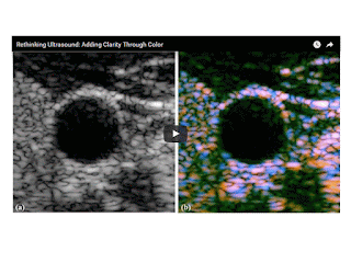
Inspired by a 19th-century set of mathematical functions, University of Rochester researcher Kevin Parker has devised a way to incorporate new color identifications in ultrasound medical images, making it easier to differentiate fine details that currently appear as indistinguishable objects in shades of gray.
The new imaging format would be especially valuable in helping physicians interpret ultrasound images of soft tissue, including muscle, glands, and organs such as the liver.
“This has been a great goal” of ultrasound research since the 1970s, said Parker, the William F. May Professor of Engineering. The technology, described in a paper in Physics in Medicine & Biology, provides more detailed soft tissue images than other attempts, based on quantitative backscattered imaging, Parker said.
Ultrasound uses pulses of high-frequency sound waves that bounce back echoes when they strike cells or arteries. These echoes are also known as scattered waves. The bouncing back gives the ultrasound image its features. With few exceptions – such as Doppler-enhanced ultrasound imaging of blood flow – those features consist of varying shades of black, white and gray reflecting different densities.
“If you look at an ultrasound image of the liver, there are so many things in there – veins, arteries, biliary ducts, liver cells, perhaps some scar tissue – and they’re all just displayed as black and white blobs,” Parker said. “If there’s a large artery, it’s easy to see the wall and the blood inside. But at the finer levels of detail, it is often impossible to tell if you’re looking at a smaller artery or 10 little cells.”
A set of mathematical functions — devised in 1890 by the great mathematician Charles Hermite of France and rarely used in engineering — provided Parker with a way to approach this problem. He came across the functions while perusing a handbook of transforms and applications, and immediately recognized that Hermite’s functions closely approximated ultrasound pulses.
“I realized if we used these, it would make our analyses of ultrasound scattering easier,” Parker said. “So now, instead of ultrasound images showing all of these tissue structures as black and white objects, we can now classify them mathematically (by their size) and assign unique colors to unique types of scatterers.”

Thứ Ba, 21 tháng 2, 2017

ARFI ĐÁNH GIÁ ĐỘ CỨNG CỦA GAN Ở BỆNH NHÂN TIỂU ĐƯỜNG TYPE 2

ARFI ĐÁNH GIÁ ĐỘ CỨNG CỦA GAN Ở BỆNH NHÂN TIỂU ĐƯỜNG TYPE 2  
NGUYỄN THỊ HỒNG ANH - NGUYỄN THIỆN HÙNG - PHAN THANH HẢI
TRUNG TÂM Y KHOA MEDIC HÒA HẢO
THÀNH PHỐ HỒ CHÍ MINH
ABSTRACT:

Using ARFI SIEMENS S2000 for measuring liver stiffness to grade the degree of liver fibrosis regardless liver steatosis. 80 adult type 2 diabetic patients were performed systematically to identify those with significant liver fibrosis. A significant liver stiffness increase was found in more than 40% of DM patients.
ARFI technique is comparable with TE in liver stiffness assessment.

ĐẶT VẤN ĐỀ
Dùng siêu âm đàn hồi ARFI SIEMENS S2000 đánh giá độ cứng gan (liver stiffness)  bệnh nhân tiểu đường, từ đó lượng giá mức độ xơ hóa của gan (fibrosis). Đánh giá độ thấm mỡ của gan (liver steatosis) bằng siêu âm B-mode.

ĐỐI TƯỢNG-PHƯƠNG PHÁP
ž  Đối tượng: bệnh nhân tiểu đường trên 18 tuổi và bình giáp, không đang mang thai hay cho con bú, không bị nhiễm HBV, HCV, không bị viêm gan tự miễn hay viêm gan do thuốc, không uống rượu bia quá 20g/ngày.
ž  Phương pháp:
·         Bệnh nhân được chẩn đoán tiểu đường và thỏa các tiêu chuẩn chọn bệnh.
·         Các xét nghiệm bao gồm đường huyết lúc đói, HbA1C, bilan lipid máu (LDL-Cholesterol, Triglyceride), creatinin, AST, ALT, GGT, FT4, TSH, HBsAg, AntiHBs, AntiHCV, Công thức máu. Xét nghiêm viêm gan tự miễn được thực hiện nếu men gan cao mà không nhiễm viêm gan siêu vi (LKM-1, AMA-M2, ANA).
·         Dùng siêu âm B-mode phát hiện và phân loại gan thấm mỡ S1, S2, S3  và không thấm mỡ S0.
·         ARFI gan được thực hiện sau siêu âm B-mode.
·         Thiết kế nghiên cứu cắt ngang, mô tả.

BẢNG PHÂN LOẠI GAN THẤM MỠ (B-MODE)


KẾT QUẢ
ž  Gồm 80 trường hợp bệnh nhân tiểu đường type 2 từ 28-79 tuổi, trong đó có 27 nam. Từ tháng 11-2016 đến tháng 02-2017.
ž  Được tổng hợp trong bảng kê dưới đây.
Số ca
S0
S1
S2
S3
Tổng
F0
5
6
6
0
17
F1
4
18
6
0
28
F2
2
3
6
1
12
F3
2
4
4
1
11
F4
0
5
4
3
12
Tổng
13
36
26
5
80

BÀN LUẬN
  Kỹ thuật không bị hạn chế khi bệnh nhân bị béo phì, báng bụng, khoang liên sườn hẹp.
  Xơ hóa gan đáng kể (F2-F3): 28,75% (23 ca) ở bệnh nhân có hoặc không có gan thấm mỡ.
  Xơ hóa gan nặng (F4):  15% (12 ca) đều ở nhóm bệnh nhân có gan thấm mỡ .
  100% gan thấm mỡ nặng (S3: 5ca) có xơ hóa gan nặng/đáng kể .
  5% (4 ca) có xơ hóa gan đáng kể (F2-F3) mà không có gan thấm mỡ : ghi nhận ở bệnh nhân có thời gian mắc bệnh lâu ngày (7-15 năm).

KẾT LUẬN
  Kỹ thuật ARFI là phương tiện nhanh có giá trị giúp đánh giá độ cứng gan ở bệnh nhân tiểu đường.
  Độ tin cậy cao trong xác định bệnh nhân không có hay có xơ hóa gan mức độ trung bình (F0 đến F2) với bệnh nhân xơ hóa gan nặng (F3-F4).

TÀI LIỆU THAM KHẢO CHÍNH


1. Liver Stiffness Evaluation by Transient Elastography in Type 2 Diabetes Mellitus Patients with Ultrasound-proven Steatosis:Sporea I, Mare R, Lupușoru R, Sima A, Sirli R, Popescu A, Timar R.: J Gastrointestin Liver Dis, 2016, Vol. 25, No 2: 167-174.
2. Liver stiffness in nonalcoholic fatty liver disease: A comparison of supersonic shear imaging, FibroScan, and ARFI with liver biopsy, Christophe Cassinotto,Jérome Boursier,Victor de Lédinghen,Jérome Lebigot, Bruno Lapuyade,Paul Cales,Jean-Baptiste Hiriart,Sophie Michalak,Brigitte Le Bail,Victoire Cartier,Amaury Mouries,Frédéric Oberti,Isabelle Fouchard-Hubert,Julien Vergniol,Christophe Aubé. Hepatology, Vol 63, June 2016.
3. Principles and clinical application of ultrasound elastography for diffuse liver disease: Woo Kyoung Jeong, Hyo K. Lim, Hyoung-Ki Lee, Jae Moon Jo, Yongsoo Kim. Ultrasonography 33(3), July 2014.


Thứ Sáu, 17 tháng 2, 2017

HƯỚNG DẪN SIÊU ÂM ĐÀN HỒI GAN của HIỆP HỘI SIÊU ÂM ÂU CHÂU EFSUMB

HƯỚNG DẪN SIÊU ÂM ĐÀN HỒI GAN  của HIỆP HỘI SIÊU ÂM ÂU CHÂU EFSUMB 2017

LIVER ELASTOGRAPHY_ EFSUMB GUIDELINES 2017

PLS DOWNLOAD FULLTEXT THEO LINK


  • http://www.umbjournal.org/article/S0301-5629(15)00220-3/pdf

Thứ Bảy, 11 tháng 2, 2017

BRAIN TUMOR ELASTiCITY _ INTRAOPERATIVE SWE

 Abstract 

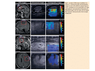
Purpose: Objective Shear wave elastography (SWE) enabled living tissue assessment of stiffness. This is routinely used for breast, thyroid and liver diseases, but there is currently no data for the brain. We aim to characterize elasticity of normal brain parenchyma and brain tumors using SWE.

Materials and Methods: Patients with scheduled brain tumor removal were included in this study. In addition to standard ultrasonography, intraoperative SWE using an ultrafast ultrasonic device was used to measure the elasticity of each tumor and its surrounding normal brain. Data were collected by an investigator blinded to the diagnosis. Descriptive statistics, box plot analysis as well as intraoperator and interoperator reproducibility analysis were also performed.

Results: 63 patients were included and classified into four main types of tumor: meningiomas, low-grade gliomas, high-grade gliomas and metastasis. Young’s Modulus measured by SWE has given new insight to differentiate brain tumors: 33.1 ± 5.9 kPa, 23.7 ± 4.9 kPa, 11.4 ± 3.6 kPa and 16.7 ± 2.5 kPa, respectively, for the four subgroups. Normal brain tissue has been characterized by a reproducible mean stiffness of 7.3 ± 2.1 kPa. Moreover, low-grade glioma stiffness is different from high-grade glioma stiffness (p = 0.01) and normal brain stiffness is very different from low-grade gliomas stiffness (p under 0.01).

Conclusion: This study demonstrates that there are significant differences in elasticity among the most common types of brain tumors. With intraoperative SWE, neurosurgeons may have innovative information to predict diagnosis and guide their resection.


Thứ Sáu, 10 tháng 2, 2017

Sir Peter Mansfield died

Sir Peter Mansfield

Professor Sir Peter Mansfield FRS,
1933-2017

Tributes have been paid to Nobel laureate Professor Sir Peter Mansfield, who has died at the age of 83.

Sir Peter pioneered the creation of Magnetic Resonance Imaging (MRI), one of the most important and revolutionary breakthroughs in modern medical science.

He was awarded the Nobel Prize for Physiology or Medicine in 2003 for his role in the development of MRI, which is used today in research, diagnosis and the treatment of millions of patients around the world.
The Sir Peter Mansfield Imaging Centre at The University of Nottingham was named in honour of his pioneering work to change the face of modern medical science.

Sir Peter Mansfield


ULTRASOUND and PULMONARY EMBOLISM

By Erik L. Ridley, AuntMinnie staff writer
February 8, 2017 -- Adding lung and venous ultrasound into the diagnostic process for pulmonary embolism (PE) can yield improved sensitivity and specificity, obviating the need for CT pulmonary angiography in many cases, according to research that will be published in the March issue of Academic Emergency Medicine.
In a multicenter study involving more than 400 patients, an Italian research team found that combining lung and venous ultrasound, Wells score, and D-dimer test results would have avoided the need to perform CT pulmonary angiography (CTPA) in half of cases with suspected PE. In comparison, a traditional approach based only on the Wells score would have bypassed CTPA in just over 25% of the patients, concluded the team led by Dr. Peiman Nazerian from Careggi University Hospital in Florence.
As a result, a diagnostic strategy that integrates clinical information, lung and venous ultrasound, and D-dimer results "may increase the performance of risk stratification and may reduce the use of CTPA in the diagnostic approach to PE, [while] still maintaining an acceptable safety profile," the authors wrote.

Thứ Sáu, 3 tháng 2, 2017

ROTATOR CUFF TEARS: U/S vs MRI


Ultrasound may unlock appendicitis treatment options

By Erik L. Ridley, AuntMinnie staff writer
February 2, 2017 -- Ultrasound may be able to differentiate between complicated and uncomplicated appendicitis, potentially helping physicians decide whether to proceed directly to an appendectomy or try antibiotics first, according to research published in the February issue of the Journal of Ultrasound in Medicine.
In a retrospective study of more than 100 patients, researchers from Stanford University found that one ultrasound finding -- the loss of the submucosal layer of the appendix -- produced 100% sensitivity and 92% specificity for complicated appendicitis.
"When ultrasound shows loss of the submucosal layer, complicated appendicitis is likely present and, therefore, surgery is likely to be necessary," senior author Dr. Eric Olcott told AuntMinnie.com. "On the other hand, when ultrasound does not show loss of the submucosal layer, antibiotics-first therapy can be considered.


"Long-axis sonogram from a 19-year-old man with gangrenous appendicitis without perforation. Image shows global loss of the submucosal layer of the appendix (A).

Thứ Năm, 2 tháng 2, 2017

THYROID ULTRASOUND, STATE of the ART, Part 1

http://www.medultrason.ro/medultrason/index.php/medultrason/article/view/980

New EFSUMB Guidelines on Liver Elastography 2017: Why and for Whom?

The new EFSUMB Guidelines on Liver Elastography 2017: why and for whom? Ioan Sporea, MD, PhD Department of Gastroenterology and Hepatology, “Victor Babes” University of Medicine and Pharmacy Timisoara, Romania

DOWNLOAD  EFSUMB Guidelines on Liver Elastography

Some years ago, during the WFUMB Meeting in Vienna in 2011, leaders in the field of ultrasound decided to issue the first European Guidelines on ultrasound based elastography. For more than a year, more than 25 experts in different fields worked together and finally the first ever EFSUMB Guidelines and Recommendations on Elastography were published in 2013 in the European Journal of Ultrasound (Ultraschall in der Medizin). This first part of the Guidelines presented data regarding the physics and technology of elastography, while the second presents the clinical applications of elastography covering 7 organs (including liver, breast, thyroid, prostate [1,2]. For many specialists in the field of ultrasound, these Guidelines were the first contact with elastography and gave them information on this new method. Following the European Guidelines, some of the National Societies issued their own Guidelines on liver elastography, starting from their large experience in the field. The Japanese Guidelines on Liver Elastography [3] and the Romanian Guidelines and Recommendations on Liver Elastography [4] were good papers to use in clinical practice. The World Federation of Ultrasound in Medicine and Biology (WFUMB) decided to issue its own Guidelines on Elastography, which were published in three parts in 2015 in the Ultrasound in Medicine and Biology journal [5-7]. These Guidelines cover to a very high level the physics of elastography as well as liver and breast elastography, enabling this method to be spread at global level.

During the last years, the development of liver elastography was impressive. Many elastographic systems appeared on the market and, in this moment, Shear Wave Elastography (SWE) is almost exclusively used for liver stiffness evaluation. SWE methods include Transient Elastography, point SWE, and 2D-SWE. Since many papers were published on this topic in the last two years, EFSUMB decided to issue new Guidelines on liver elastography. Working together for many months, a group of approximately 20 experts in the field of liver elastography succeeded to produce the new EFSUMB Guidelines on liver Elastography 2017 (EFSUMB Guidelines and Recommendations on the Clinical Use of Liver Ultrasound Elastography, update 2017). This paper was sent for publication in Ultraschall in der Medizin and will appear very soon.

 The first question is why do we need new EFSUMB Guidelines on liver elastography? The answer is because the body of evidence accumulated in the last 3-4 years on this topic is so abundant that the new information must refined and send to the specialists in the field. Secondly, because the practical experience on liver elastography is now extensive and allows pertinent recommendations. As compared with the first EFSUMB Guidelines, the second edition presents data on liver elastography not only with Transient Elastography, but also the experience with point SWE (especially VTQ) and 2D-SWE (especially SSI). The first section of the paper presents the physics and the systems used in liver elastography and is continued with practical recommendations on how to use this method. 

The second question is to whom are these Guidelines addressed to. First of all, to anyone who likes to know what is new in the field of medicine. Secondly to specialists who work in the field of hepatology, because in the last years liver stiffness evaluation using elastography replaced in many cases liver biopsy. In many countries, including Romania, liver elastography replaced quite  Ioan Sporea. The new EFSUMB Guidelines on Liver Elastography 2017: why and for whom? completely liver biopsy (especially in C chronic viral hepatitis). Ultrasound based elastography of the liver is performed by gastroenterologists and hepatologists, internal medicine specialist, and by radiologists. If in the USA, Magnetic Resonance Elastography (MRE) is continuously developing, in Europe liver elastography is performed quite exclusively by ultrasound. 

Considering the results of published papers and metaanalyses, the new EFSUMB Guidelines publish cut-off values for different liver elastographic methods, in different chronic liver diseases (such as chronic viral hepatitis, alcoholic and non-alcoholic steatohepatitis, and others). Limitations and tricks of these methods are presented. Finally, the new EFSUMB Guidelines on Liver Elastography are welcome for everybody. It is a well done material, written by the most experienced European specialists in liver elastography, very useful for practitioners.