Tổng số lượt xem trang

Thứ Tư, 14 tháng 12, 2016

CỠ MẪU: Huyền thoại con số 30

Download theo link:Cỡ mẫu
http://vienthongke.vn/attachments/article/1644/Bai3So%205_2012.pdf

Thứ Tư, 23 tháng 11, 2016

ARFI of FATTY LIVER and DIABETIC LIVER @ MEDIC CENTER

ABSTRACT:

Using Fibroscan 502 TOUCH and ARFI SIEMENS S2000 for evaluation of fatty liver infiltration and diabetic liver on 84 patients we realized that CAP could detect quantitatively fatty composition with high values of CAD in liver, but Fibroscan and ARFI had low values of SWE speed.

ĐẶT VẤN ĐỀ

Dùng siêu âm đàn hồi gồm Fibroscan 502 Touch và ARFI SIEMENS S 2000 cùng lúc để đánh giá gan thấm mỡ (fatty liver infiltration)  và gan bệnh nhân tiểu đường để khảo sát gan thấm mỡ và gan bệnh nhân tiểu đường ở người Việt nam.

Chúng tôi thực hiện 2 kỹ thuật Fibroscan (EchoSense) và ARFI Siemens cùng lúc trên 84 bệnh nhân gan thấm mỡ với hơn 1/3 bệnh nhân tiểu đường type 2, tuổi từ 20-55.

PHƯƠNG PHÁP-ĐỐI TƯỢNG

Thiết kế nghiên cứu cắt ngang, mô tả. 


Dùng siêu âm B-mode phát hiện và phân loại gan thấm mỡ  S1, S2, S3, và dựa vào thông số hóa sinh (triglycerid, cholesterol, lipid máu, ALT, HbA1c, tiểu cầu). Sau đó bệnh nhân được làm FibroScan và định lượng CAP [ độ nhiễm mỡ gan dựa trên “thông số giảm âm được kiểm soát” (Controlled Attenuation Parameter: CAP) bằng cách tính toán từ tín hiệu siêu âm trong phép đo độ cứng].Phần khảo sát ARFI gan thấm mỡ được làm sau phần khảo sát gan bằng B-mode.

KẾT QUẢ của thăm dò như trong bảng sau:


BÀN LUẬN


tương hợp không đồng đều giữa đánh giá siêu âm B-mode (S1,2,3) C A P của Fibroscan.


KẾT LUẬN:

1/ CAP của FIBROSCAN giúp định lượng mỡ trong gan  không cần phải sinh thiết.
2/ Tích tụ mỡ trong gan vẻ không làm tăng độ cứng   gan do đó không làm tăng tốc độ đàn hồi sóng biến dạng.

Đây chỉ là số liệu ban đầu, cần thực hiện với mẩu chọn lớn hơn.  CAP của FibroScan  giúp đánh giá định lượng mức độ gan thấm mỡ mà không phải sinh thiết gan, trong khi transient elastography của Fibroscan và máy siêu âm Siemens khảo sát độ đàn hồi của gan thấm mỡ với kết quả tương đương nhau, với tốc độ sóng biến dạng trong gan thấm mỡ không tăng.

TÀI LIỆU THAM KHẢO CHÍNH
Geoffrey C. Farrell, Arthur J. McCullough and Christopher P. Day: Non-Alcoholic Fatty liver Disease, Wiley-Blackwell, 2013.
Ioan Sporea, Ruxandra Mare, Raluca Lupușoru, Alexandra Sima, Roxana Șirli, Alina Popescu, Romulus Timar: Liver Stiffness Evaluation by Transient Elastography in Type 2 Diabetes Mellitus Patients with Ultrasound-proven Steatosis, J Gastrointestin Liver Dis, June 2016 Vol. 25 No 2: 167-174.
Yin-Yan Li, Xue-Mei Wang, Yi-Xia Zhang, Guo-Cheng Ou:Ultrasonic elastography in clinical quantitative assessment of fatty liver, World J Gastroenterol 2010 October 7; 16(37): 4733-4737.

Thứ Ba, 22 tháng 11, 2016

Diabetes Type 2 và SIÊU ÂM Elastography @ MEDIC HOA HAO CENTER

Ultrasound and transient elastography: Screening exams for type 2 diabetes mellitus patients

By Staff News Brief
Liver stiffness assessment should be routinely performed on individuals with Type 2 diabetes mellitus (DM), as these patients are susceptible to developing nonalcoholic fatty liver disease (NAFLD). Gastroenterologists from Romania recommend using ultrasound and elastography to identify and assess the severity of liver steatosis and fibrosis among their patients. Their study findings were published in the June 2016 issue of the Journal of Gastrointestinal Liver Disease.
Diabetes and obesity have reached epidemic proportions in Western societies, and the number of patients afflicted by either condition continues to rise. A large percentage of patients with DM are obese and may develop liver steatosis, the abnormal retention of triglycerides and lipids in liver cells. Patients who have NAFLD may develop nonalcoholic steatohepatis and subsequently liver cirrhosis.
Lead author Ioan Sporea, MD, of the Department of Gastroenterology and Hepatology of the Victor Babeş, a University of Medicine and Pharmacy in Timişoara, and colleagues, state that NAFLD will become one of the most important problems to confront hepatologists as a leading cause of liver cirrhosis. Biopsy, the gold standard for diagnosis, may become impractical to perform on all patients. Because inflammation can differentiate severe from simple steatosis, the authors conducted a study of nearly 300 patients to assess the extent and severity of liver steatosis using ultrasound, and of liver fibrosis using transient elastography.

(A) mild steatosis, (B) moderate steatosis, (C) severe steatosis
The 298 patients with Type 2 DM were enrolled in the study during a six month period in 2013. An additional 109 healthy volunteers and 43 hospitalized patients without liver disease completed the study cohort. Based on ultrasound findings, they were grouped into one of four steatosis categories ranging from none to severe. Liver stiffness as a marker of fibrosis was evaluated by transient elastography.
A total of 260 patients (87.2%) were identified as having steatosis: 44 patients had severe and 118 patients had moderate steatosis. 56, or 18.8% patients diagnosed with NAFLD had significant fibrosis. Liver stiffness suggestive of cirrhosis was identified in 41 (13.8%) patients. An evaluation to determine if risk of fibrosis increased in NAFLD patients who were obese and had type 2 DM determined that obesity, steatosis, higher alanine aminotransferase, and hypertriglyceridemia were independently associated with liver stiffness values. There were no significant differences between the mean liver stiffness value in obese, underweight, or normal weight individuals in the control group.
The authors recommend that ultrasound be used in daily practice to screen diabetic patients for steatosis because of its availability, low cost, rapid exam time of approximately five minutes, and diagnostic accuracy. They also recommend the use of ultrasound-based elastographic techniques; ie, transient elastography for liver fibrosis assessment.

REFERENCE

  1. Sporea I, Mare R, Lupuşoru R, et al. Liver stiffness evaluation by transient elastography in type 2 diabetes mellitus patients with ultrasound-proven steatosis. J Gastrointestin Liver Dis. 2016 25;2: 167-174.

Ultrasound and transient elastography: Screening exams for type 2 diabetes mellitus patients.  Appl Radiol. 
By Staff News Brief| July 18, 2016
Categories:  Section

About the Author

Staff News Brief

Staff News Brief

NGUYÊN LÝ và ỨNG DỤNG LÂM SÀNG SIÊU ÂM ĐÀN HỒI TRONG BỆNH LÝ GAN LAN TỎA

DOWNLOAD HERE

Abstract

Accurate assessment of the degree of liver fibrosis is important for estimating prognosis and deciding on an appropriate course of treatment for cases of chronic liver disease (CLD) with various etiologies. Because of the inherent limitations of liver biopsy, there is a great need for non-invasive and reliable tests that accurately estimate the degree of liver fibrosis. Ultrasound (US) elastography is considered a non-invasive, convenient, and precise technique to grade the degree of liver fibrosis by measuring liver stiffness. There are several commercial types of US elastography currently in use, namely, transient elastography, acoustic radiation force impulse imaging, supersonic shear-wave imaging, and real-time tissue elastography. Although the low reproducibility of measurements derived from operator-dependent performance remains a significant limitation of US elastography, this technique is nevertheless useful for diagnosing hepatic fibrosis in patients with CLD. Likewise, US elastography may also be used as a convenient surveillance method that can be performed by physicians at the patients’ bedside to enable the estimation of the prognosis of patients with fatal complications related to CLD in a non-invasive manner.

Thứ Tư, 5 tháng 10, 2016

Basic Concepts of Doppler Frequency Spectrum Analysis and Ultrasound Blood Flow Imaging

DOWNLOAD FULLTEXT THEO LINK

John S. Pellerito, MD, FACR, FSRU, FAIUM , Joseph F. Polak, MD, MPH


Góc Doppler = là góc tạo bởi phương ngắm hay đường ngắm Doppler [Doppler line 

of sight] và trục dòng máu chảy. Xem minh họa dưới đây.


Thứ Ba, 30 tháng 8, 2016

AIUM PRACTICE GUIDELINE on J U M 9-2016

  • AIUM PRACTICE GUIDELINE

    • AIUM Practice Parameter for the Performance of an Ultrasound Examination of the Extracranial Cerebrovascular System

    Journal of Ultrasound in Medicine September 2016 35:1-11;doi:10.7863/ultra.35.9.1
  • AIUM PRACTICE GUIDELINE

      AIUM Practice Parameter for the Performance of Ultrasound Vascular Mapping for Preoperative Planning of Dialysis Access

    Journal of Ultrasound in Medicine September 2016 35:1-10;doi:10.7863/ultra.35.9.1-a
  • AIUM PRACTICE GUIDELINE

      AIUM Practice Parameter for the Performance of an Ultrasound Examination of the Neonatal and Infant Spine

    Journal of Ultrasound in Medicine September 2016 35:1-11;doi:10.7863/ultra.35.9.1-b
  • AIUM PRACTICE GUIDELINE

      AIUM Practice Parameter for the Performance of a Thyroid and Parathyroid Ultrasound Examination

    Journal of Ultrasound in Medicine September 2016 35:1-11;doi:10.7863/ultra.35.9.1-c
  • AIUM PRACTICE GUIDELINE

      AIUM Practice Parameter for the Performance of Selected Ultrasound-Guided Procedures

    Journal of Ultrasound in Medicine September 2016 35:1-40;doi:10.7863/ultra.35.9.1-d
5


Thứ Bảy, 13 tháng 8, 2016

VIÊM RUỘT DƯ VÀ K DẠ DÀY

DOWNLOAD appendicitis and gastric cancer


ABSTRACT Primary non-carcinoid adenocarcinoma of the appendix is rare. Likewise, distant metastatisis of another organ or system cancer is even more rare. Generally, gastric adenocarcinoma may clinically be detected while it is spreaded. A 31-year-old man who had no specific medical history was admitted to the clinic with complaint of right lower abdominal quadrant pain and rebound tenderness over McBurney’s point. Laparotomy was performed for a diagnosis of acute appendicitis. Histopathological examination revealed an obstruction of the appendicular lumen due to adenocarcinoma metastasis. Correspondingly, the patient was re-evaluated to detect the primary malignancy focus and underwent inoperable gastric adenocarcinoma diagnosis. As our knowledge, there is only one other example in the literature; a patient with undiagnosed gastric cancer who had an acute appendicitis as the first clinical manifestation of an upper gastrointestinal malignancy. Thus we want to share this unusual, interesting and complicated case.


Key words: Acute appendicitis, Gastric adenocarcinoma, Distant metastasis, Diagnostic laparoscopy  

Thứ Bảy, 6 tháng 8, 2016

SIÊU ÂM VÀ CAO HUYẾT ÁP BỆNH NHÂN TIỂU ĐƯỜNG


Sounding out high blood pressure in diabetics

July 28, 2016
http://medicalxpress.com/news/2016-07-high-blood-pressure-diabetics.html
Sounding-out high blood pressure in diabetics
Applying ultrasound could be a cheap, non-invasive method to treat treatment-resistant hypertension in diabetics. Credit: Tohoku University
Blood pressure can significantly drop by applying 20 minutes of ultrasound to the forearm of type II diabetes patients with treatment-resistant hypertension, according to research from Japan's Tohoku University.

Thứ Sáu, 8 tháng 7, 2016

Esophageal Varices and SWE

By Erik L. Ridley, AuntMinnie staff writer
July 7, 2016 -- Shear-wave elastography can noninvasively predict the presence of esophageal varices in patients with the first stage of liver cirrhosis, and bests other measures for spotting high-risk varices, according to research published in the July issue of the Journal of Ultrasound in Medicine.

Thứ Hai, 27 tháng 6, 2016

CHUẨN BỊ THAM GIA HỘI NGHỊ CHẨN ĐOÁN HÌNH ẢNH TOÀN QUỐC LẦN 18 19-20/8 ĐÀ NẴNG

Gồm 8 bài báo cáo về siêu âm:

1/ ARFI U VÚ LÀNH TÍNH VÀ ÁC TÍNH, BS VÕ NGUYỄN THỤC QUYÊN, BS LÊ THANH LIÊM, BS PHAN THANH HẢI PHƯỢNG, BS PHAN THANH HẢI

2/ TÚI GIẢ PHÌNH TĨNH MẠCH THẬN TRÁI, CHẨN ĐOÁN VÀ ĐIỀU TRỊ, BS NGUYỄN NGHIỆP VĂN

3/ GIẢNG DẠY GIẢI PHẪU HỌC BẰNG SIÊU ÂM: KINH NGHIỆM BAN ĐẦU, BS NGUYỄN THIỆN HÙNG-BS PHAN THANH HẢI

4/ SMI TRONG CHÂN DOÁN U GAN, BS NGUYEN NGHIEP VAN , BS PHAN THANH HAI PHUONG, BS PHAN THANH HAI

5/ KHẢO SÁT VI MẠCH SMI NHÂN GIÁP LÀNH TÍNH VÀ ÁC TÍNH, BS LÊ TỰ PHÚC

6/ U tụy đặc giả nhú ở trẻ em (case report), BS LÊ THANH LIÊM
7/ SIÊU ÂM PHÁT HIỆN GÃY XƯƠNG SƯỜN, BS LÊ THANH LIÊM.
8/ ARFI THẬN BẾ TẮC DO SỎI VÀ DO HẸP KHÚC NỐI, BS TRẦN NGÂN CHÂU.

Download chuong trinh hoi nghi CDHA chi tiet

CHUONG TRINH HOi NGHI CDHA 2016
http://vcr2016.com.vn/Content.aspx?cid=53

Thứ Ba, 7 tháng 6, 2016

Vai trò siêu âm SMI trong chẩn đoán u gan và theo dỏi điều tri

DOWNLOAD theo link

vai-tr-siu-m-smi-trong-chn-on-u-gan-va-theo-di-iu-tr
vai tro sieu am SMI trong chan doan u gan va theo doi dieu tri


Vai Trò Của Siêu Âm Tạo hình Vi mạch SMI
(Superb Micro-Vascular Imaging)
Trong Bệnh Lý Mạch Máu

I.                   Đặt Vấn Đề:

Số lượng bệnh nhân khám siêu âm mạch máu ngày càng nhiều. phát hiện bệnh lý mạch máu và đánh giá huyết động chính xác là cần thiết cho lập kế hoạch điều trị.
Với những tổn thương hẹp động mạch hay huyết khối tĩnh mạch, vai trò của siêu âm Doppler trong khảo sát tái tưới máu của huyết khối và đánh giá tính không ổn định của mảng xơ vữa được đặt ra. Điều này đòi hỏi phát hiện những mạch máu nhỏ, lưu lượng dòng chảy thấp mà không sử dụng chất cản âm.
Với các mode siêu âm màu đã có ở các thế hệ máy trước đây như siêu âm màu thường qui (Color Doppler), siêu âm màu năng lượng (Power Doppler) và siêu âm màu dòng chảy động (Advance Dynamic flow) đã giúp đánh giá sự phân bố mạch máu ngoại biên. Tuy nhiên hạn chế của các mode siêu âm màu kể trên là khó khảo sát những mạch máu nhỏ có lưu lượng dòng chảy thấp và dễ bị xảo ảnh do chuyển động (motion artifact) gây ra.

II.                Kỹ Thuật SMI (Superb Micro – Vascular Imaging)

Với kỹ thuật SMI đã khảo sát được những mạch máu rất nhỏ, có dòng chảy thấp, tỉ lệ khung ảnh và độ nét cao, giảm được xảo ảnh gây ra chuyển động mô, có thể tạo hình mạch máu nhỏ hơn với tốc độ dòng chảy thấp hơn mà không dùng chất cản âm. Ưu điểm này hứa hẹn là  kỹ thuật mới vô cùng hữu ích trong việc đánh giá sự phân bố mạch máu trong các tổn thương bướu, đánh giá tái tưới máu huyết khối, đánh giá tân sinh mạch trong mảng xơ vữa không ổn định.
SMI phân biệt được tín hiệu màu ngoại lai (do motion artifact), lọc chính xác tín hiệu mạch máu rất nhỏ.

SMI bao gồm 2 mode:
           SMI màu (cSMI): mạch máu tín hiệu thấp phổ màu.
           SMI trắng-đen (mSMI): hình ảnh mạch máu độ nhạy cao hơn, nổi bật hơn do xóa tín hiệu nền.

III.             Ứng Dụng Của SMI

1.     Khảo sát tân sinh mạch trong mảng xơ vữa động mạch cảnh

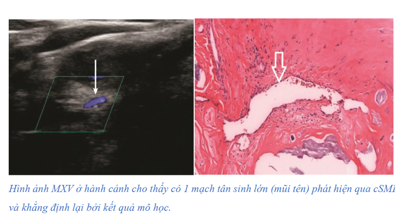
Mảng xơ vữa (MXV) động mạch cảnh gây ra 30% các ca đột quỵ, không phải do hẹp động mạch nhưng do bong MXV không ổn định. Y văn khuyến cáo nên xem xét tính không ổn định của MXV khi đánh giá nguy cơ đột quỵ. Ngoài đánh giá độ hẹp lòng mạch cảnh, cần xem xét các yếu tố khác như xuất huyết trong MXV, tân sinh mạch trong mảng và lớp vỏ xơ mỏng có thể giúp cải thiện phân tầng nguy cơ và kết cục của người bệnh.
Các kỹ thuật siêu âm không cản quang hiện nay chưa tối ưu trong đánh giá MXV không ổn định. Ngay cả CTA chỉ đánh giá được độ hẹp, mà không đánh giá được MXV không ổn định. Kỹ thuật SMI có thể nhận diện tín hiệu chuyển động ngoài dòng máu và tách khỏi tín hiệu Doppler. Đặc tính này làm SMI thu nhận được hình các dòng chảy chậm mà bị loại bỏ bởi độ lọc thành của Doppler thường qui. 


0
1
2
3
Siêu âm

Không có dòng chảy bên trong MXV
Dòng chảy nhỏ chỉ nhìn thấy trên SMI
Nhiều vị trí có dòng chảy nhìn thấy bằng SMI và dòng chảy nhỏ nhìn thấy trên Doppler năng lượng
Dòng chảy ưu thế nhìn thấy trên SMI và Doppler năng lượng
Bệnh học
Không có dòng chảy bên trong MXV
Vài dòng chảy nhỏ được nhìn thấy trong MXV
Mạch máu nhỏ và 1-2 mạch máu lớn hơn trong MXV
Nhiều mạch máu lớn và nhỏ trong MXV
Bảng  phân độ tân sinh mạch theo hình ảnh siêu âm và kết quả mô học của MXV.


Khảo sát mốt số trường hợp tại Medic
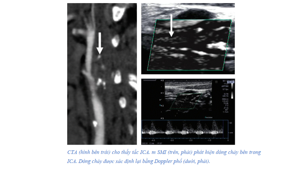


2.      Khảo sát tái tưới máu trong huyết khối, đánh giá hiệu quả xơ hóa tĩnh mạch hiển lớn sau can thiệp nội mạch


Với tính năng làm giảm xảo ảnh do chuyển động, và cho phép tạo hình dòng chảy tốc độ thấp, độ tương phản âm giữa huyết khối và dòng chảy xung quanh giúp nhìn rõ huyết khối tĩnh mạch khi làm trên SMI so với B-mode và Doppler màu truyền thống. SMI là một phương tiện hữu ích đánh gía huyết khối, nhất là trong giai đoạn sớm sau khi hình thành huyết khối, để phác họa kích thước và chiều dài huyết khối. Với huyết khối đang điều trị, SMI giúp đánh giá dòng chảy tái thông với độ nhạy tốt hơn so Doppler màu qui ước.


Khảo sát tại Medic:


   Đánh giá huyết khối bám thành trong phình động mạch chủ bụng và biến chứng rò sau stent graft động mạch chủ bụng.


IV.          Kết Luận:

Thách thức về mặt lâm sàng tồn tại trong việc phát hiện những mạch máu nhỏ, lưu lượng dòng chảy thấp mà không sử dụng chất cản âm.  Trong điều kiện đó, kỹ thuật SMI ra đời, với những tính năng như tạo được hình ảnh dòng chảy tốc độ thấp, độ phân giải cao, hạn chế tốt xảo ảnh do chuyển động, tốc độ khung hình cao. Nhất là kỹ thuật này có thể tạo hình mạch máu nhỏ hơn với tốc độ dòng chảy thấp hơn mà không dùng chất cản âm. Với những ưu điểm này, kỹ thuật mới hứa hẹn nhiều  tiềm năng trong sử dụng đánh giá tái tưới máu.

Tài liệu tham khảo


1. Tokodai K, Miyagi S, Nakanishi C, Hara Y, Nakanishi W, Miyazawa K, Shimizu K, Goto M, Kamei T, Unno M. The utility of superb microvascular imaging for monitoring low-velocity venous flow following pancreas transplantation: report of a case. J Med Ultrason (2001). 2018 Jan;45(1):171-174

2. Oura KKato TOhba HTerayama Y. Evaluation of Intraplaque Neovascularization Using Superb Microvascular Imaging and Contrast-Enhanced Ultrasonography. J Stroke Cerebrovasc Dis. 2018 Sep; 27(9):2348-2353

3. Hagiwara YSasaki RShimizu TSoga KHatada CMiyauchi MOkamura TSakurai MAkiyama HHasegawa Y. The utility of superb microvascular imaging for the detection of deep vein thrombosis. J Med Ultrason (2001). 2018 Oct;45(4):665-669

4. Canon Medical.  Ultrasound Clinical Case Study Portal Vein Thrombosis
5. Marcin Gabriel, Jolanta Tomczak, Magdalena Snoch-Ziółkiewicz, Łukasz Dzieciuchowicz, Ewa Strauss, Katarzyna Pawlaczyk, Dorota Wojtusik, Grzegorz Oszkinis. Superb Micro-vascular Imaging (SMI): a Doppler ultrasound technique with potential to identify, classify, and follow up endoleaks in patients after Endovascular Aneurysm Repair (EVAR)Abdom Radiol (NY) 2018; 43(12): 3479–3486